 Movie
Movie Controller

[English] 日本語
 Yorodumi
- PDB-2wh0: Recognition of an intrachain tandem 14-3-3 binding site within pr... -
+ Open data

- Basic information
| Entry | Database: PDB / ID: 2wh0 | ||||||
|---|---|---|---|---|---|---|---|
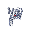
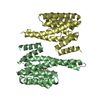
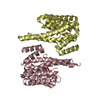
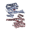
| Title | Recognition of an intrachain tandem 14-3-3 binding site within protein kinase C epsilon | ||||||
 Components Components |
| ||||||
 Keywords Keywords | SIGNALING PROTEIN / TANDEM BINDING / PHOSPHOPROTEIN / 14-3-3 / CYTOPLASM / ACETYLATION / PKC EPSILON | ||||||
| Function / homology |  Function and homology information Function and homology informationethanol binding / TRAM-dependent toll-like receptor 4 signaling pathway / diacylglycerol-dependent, calcium-independent serine/threonine kinase activity / negative regulation of sodium ion transmembrane transport / toxin catabolic process / diacylglycerol-dependent serine/threonine kinase activity / DAG and IP3 signaling / protein kinase C / synaptic target recognition / Golgi reassembly ...ethanol binding / TRAM-dependent toll-like receptor 4 signaling pathway / diacylglycerol-dependent, calcium-independent serine/threonine kinase activity / negative regulation of sodium ion transmembrane transport / toxin catabolic process / diacylglycerol-dependent serine/threonine kinase activity / DAG and IP3 signaling / protein kinase C / synaptic target recognition / Golgi reassembly / intermediate filament cytoskeleton / Effects of PIP2 hydrolysis / NOTCH4 Activation and Transmission of Signal to the Nucleus / respiratory system process / establishment of Golgi localization / tube formation / regulation of synapse maturation / Rap1 signalling / positive regulation of fibroblast migration / positive regulation of actin filament polymerization / negative regulation of protein localization to nucleus / KSRP (KHSRP) binds and destabilizes mRNA / GP1b-IX-V activation signalling / positive regulation of wound healing / positive regulation of cytokinesis / Fc-gamma receptor signaling pathway involved in phagocytosis / signaling receptor activator activity / positive regulation of epithelial cell migration / Regulation of localization of FOXO transcription factors / Interleukin-3, Interleukin-5 and GM-CSF signaling / actin monomer binding / phosphoserine residue binding / Activation of BAD and translocation to mitochondria / Role of phospholipids in phagocytosis / regulation of ERK1 and ERK2 cascade / xenobiotic catabolic process / lung development / SARS-CoV-2 targets host intracellular signalling and regulatory pathways / cellular response to glucose starvation / Chk1/Chk2(Cds1) mediated inactivation of Cyclin B:Cdk1 complex / SARS-CoV-1 targets host intracellular signalling and regulatory pathways / RHO GTPases activate PKNs / positive regulation of superoxide anion generation / lipopolysaccharide-mediated signaling pathway / negative regulation of TORC1 signaling / 14-3-3 protein binding / Transcriptional and post-translational regulation of MITF-M expression and activity / negative regulation of protein ubiquitination / negative regulation of innate immune response / SHC1 events in ERBB2 signaling / hippocampal mossy fiber to CA3 synapse / cell periphery / regulation of actin cytoskeleton organization / TP53 Regulates Metabolic Genes / positive regulation of protein localization to plasma membrane / Translocation of SLC2A4 (GLUT4) to the plasma membrane / regulation of protein stability / protein sequestering activity / Deactivation of the beta-catenin transactivating complex / Negative regulation of NOTCH4 signaling / enzyme activator activity / intracellular protein localization / melanosome / G alpha (z) signalling events / angiogenesis / protein phosphatase binding / blood microparticle / DNA-binding transcription factor binding / vesicle / transmembrane transporter binding / protein phosphorylation / protein kinase activity / cell adhesion / intracellular signal transduction / cadherin binding / protein domain specific binding / protein serine kinase activity / cell division / focal adhesion / protein serine/threonine kinase activity / apoptotic process / ubiquitin protein ligase binding / negative regulation of apoptotic process / protein kinase binding / perinuclear region of cytoplasm / Golgi apparatus / glutamatergic synapse / negative regulation of transcription by RNA polymerase II / enzyme binding / signal transduction / endoplasmic reticulum / mitochondrion / : / RNA binding / extracellular exosome / nucleoplasm / zinc ion binding / ATP binding / identical protein binding / nucleus Similarity search - Function | ||||||
| Biological species |  HOMO SAPIENS (human) HOMO SAPIENS (human) | ||||||
| Method |  X-RAY DIFFRACTION / MOLECULAR REPLACEMENT / Resolution: 2.25 Å X-RAY DIFFRACTION / MOLECULAR REPLACEMENT / Resolution: 2.25 Å | ||||||
 Authors Authors | Kostelecky, B. / Saurin, A.T. / Purkiss, A. / Parker, P.J. / McDonald, N.Q. | ||||||
 Citation Citation | Journal: Embo Rep. / Year: 2009 Title: Recognition of an Intra-Chain Tandem 14-3-3 Binding Site within Pkc Epsilon. Authors: Kostelecky, B. / Saurin, A.T. / Purkiss, A. / Parker, P.J. / Mcdonald, N.Q. | ||||||
| History |
|
- Structure visualization
| Structure viewer | Molecule: MolmilJmol/JSmol |
|---|
- Downloads & links
-Download
| PDBx/mmCIF format | 2wh0.cif.gz | 180.3 KB | Display | PDBx/mmCIF format |
|---|---|---|---|---|
| PDB format | pdb2wh0.ent.gz | 141.4 KB | Display | PDB format |
| PDBx/mmJSON format | 2wh0.json.gz | Tree view | PDBx/mmJSON format | |
| Others |  Other downloads Other downloads |
-Validation report
| Arichive directory | https://data.pdbj.org/pub/pdb/validation_reports/wh/2wh0ftp://data.pdbj.org/pub/pdb/validation_reports/wh/2wh0 | HTTPS FTP |
|---|
-Related structure data
| Related structure data |  1qjbS S: Starting model for refinement |
|---|---|
| Similar structure data |


-Links
 PDBj
PDBj


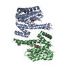
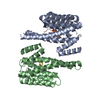
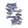
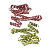
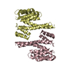
- Assembly
| Deposited unit | 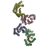
| ||||||||||||||||
|---|---|---|---|---|---|---|---|---|---|---|---|---|---|---|---|---|---|
| 1 | 
| ||||||||||||||||
| 2 | 
| ||||||||||||||||
| Unit cell |
| ||||||||||||||||
| Noncrystallographic symmetry (NCS) | NCS oper:
|
-Components
| #1: Protein | Mass: 27777.092 Da / Num. of mol.: 4 Source method: isolated from a genetically manipulated source Source: (gene. exp.) HOMO SAPIENS (human) / Plasmid: PACYC / Production host:  #2: Protein/peptide | Mass: 3728.884 Da / Num. of mol.: 2 / Fragment: PKC EPSILON V3-DERIVED PEPTIDE, RESIDUES 342-372 / Source method: obtained synthetically / Details: SYNTHETIC FRAGMENT OF PKC EPSILON V3 REGION / Source: (synth.) HOMO SAPIENS (human) / References: UniProt: Q02156, protein kinase C#3: Chemical | ChemComp-PGE / |   Mass: 150.173 Da / Num. of mol.: 1 / Source method: obtained synthetically / Formula: C6H14O4 Mass: 150.173 Da / Num. of mol.: 1 / Source method: obtained synthetically / Formula: C6H14O4#4: Chemical | ChemComp-CA / |   Mass: 40.078 Da / Num. of mol.: 1 / Source method: obtained synthetically / Formula: Ca Mass: 40.078 Da / Num. of mol.: 1 / Source method: obtained synthetically / Formula: Ca#5: Water | ChemComp-HOH / |  Mass: 18.015 Da / Num. of mol.: 64 / Source method: isolated from a natural source / Formula: H2O Mass: 18.015 Da / Num. of mol.: 64 / Source method: isolated from a natural source / Formula: H2OHas protein modification | Y | |
|---|
-Experimental details
-Experiment
| Experiment | Method: X-RAY DIFFRACTION / Number of used crystals: 1 |
|---|
- Sample preparation
| Crystal | Density Matthews: 2.58 Å3/Da / Density % sol: 52.3 % / Description: NONE |
|---|---|
| Crystal grow | pH: 7.4 Details: 16 MG/ML PROTEIN 9% (W/V) POLYETHYLENE GLYCOL 3350, 25 MM CALCIUM ACETATE, 25 MM SODIUM FLUORIDE, pH 7.4 |
-Data collection
| Diffraction | Mean temperature: 100 K |
|---|---|
| Diffraction source | Source: ROTATING ANODE / Type: RIGAKU MICROMAX-007 HF / Wavelength: 1.5418 |
| Detector | Type: MARRESEARCH / Detector: IMAGE PLATE / Date: Aug 13, 2008 / Details: OSMIC MIRRORS |
| Radiation | Protocol: SINGLE WAVELENGTH / Monochromatic (M) / Laue (L): M / Scattering type: x-ray |
| Radiation wavelength | Wavelength: 1.5418 Å / Relative weight: 1 |
| Reflection twin | Operator: h,-k,-l / Fraction: 0.441 |
| Reflection | Resolution: 2.25→24.5 Å / Num. obs: 56277 / % possible obs: 99.4 % / Observed criterion σ(I): 3.9 / Redundancy: 4.9 % / Biso Wilson estimate: 39.6 Å2 / Rmerge(I) obs: 0.1 / Net I/σ(I): 19.4 |
| Reflection shell | Resolution: 2.25→2.45 Å / Redundancy: 4.8 % / Rmerge(I) obs: 0.41 / Mean I/σ(I) obs: 3.9 / % possible all: 82.4 |
- Processing
| Software |
| |||||||||||||||||||||||||||||||||||||||||||||||||||||||||||||||||||||||||||||||||||||||||||||||||||||||||||||||||||||||||||||||||||||||||||||||||||
|---|---|---|---|---|---|---|---|---|---|---|---|---|---|---|---|---|---|---|---|---|---|---|---|---|---|---|---|---|---|---|---|---|---|---|---|---|---|---|---|---|---|---|---|---|---|---|---|---|---|---|---|---|---|---|---|---|---|---|---|---|---|---|---|---|---|---|---|---|---|---|---|---|---|---|---|---|---|---|---|---|---|---|---|---|---|---|---|---|---|---|---|---|---|---|---|---|---|---|---|---|---|---|---|---|---|---|---|---|---|---|---|---|---|---|---|---|---|---|---|---|---|---|---|---|---|---|---|---|---|---|---|---|---|---|---|---|---|---|---|---|---|---|---|---|---|---|---|---|
| Refinement | Method to determine structure: MOLECULAR REPLACEMENT Starting model: PDB ENTRY 1QJB Resolution: 2.25→24.46 Å / σ(F): 1.99 / Phase error: 37.54 / Stereochemistry target values: TWIN_LSQ_F / Details: TWINNED REFINEMENT
| |||||||||||||||||||||||||||||||||||||||||||||||||||||||||||||||||||||||||||||||||||||||||||||||||||||||||||||||||||||||||||||||||||||||||||||||||||
| Solvent computation | Shrinkage radii: 0.9 Å / VDW probe radii: 1.11 Å / Solvent model: FLAT BULK SOLVENT MODEL / Bsol: 39.57 Å2 / ksol: 0.35 e/Å3 | |||||||||||||||||||||||||||||||||||||||||||||||||||||||||||||||||||||||||||||||||||||||||||||||||||||||||||||||||||||||||||||||||||||||||||||||||||
| Displacement parameters | Biso mean: 34.93 Å2
| |||||||||||||||||||||||||||||||||||||||||||||||||||||||||||||||||||||||||||||||||||||||||||||||||||||||||||||||||||||||||||||||||||||||||||||||||||
| Refinement step | Cycle: LAST / Resolution: 2.25→24.46 Å
| |||||||||||||||||||||||||||||||||||||||||||||||||||||||||||||||||||||||||||||||||||||||||||||||||||||||||||||||||||||||||||||||||||||||||||||||||||
| Refine LS restraints |
| |||||||||||||||||||||||||||||||||||||||||||||||||||||||||||||||||||||||||||||||||||||||||||||||||||||||||||||||||||||||||||||||||||||||||||||||||||
| LS refinement shell |
|
