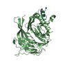
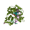
 Movie
Movie Controller


+ Open data

- Basic information
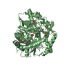
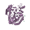
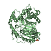
| Entry | Database: PDB / ID: 5wvr | ||||||
|---|---|---|---|---|---|---|---|
| Title | Crystal structure of Osh1 ORD domain in complex with cholesterol | ||||||
 Components Components | KLLA0C04147p | ||||||
 Keywords Keywords | LIPID BINDING PROTEIN / oxysterol binding / lipid transfer / cholesterol | ||||||
| Function / homology |  Function and homology information Function and homology informationsterol transfer activity / sterol binding / perinuclear endoplasmic reticulum / maintenance of cell polarity / piecemeal microautophagy of the nucleus / exocytosis / endocytosis / nuclear envelope / plasma membrane / cytosol Similarity search - Function | ||||||
| Biological species |  Kluyveromyces lactis (yeast) Kluyveromyces lactis (yeast) | ||||||
| Method |  X-RAY DIFFRACTION / SYNCHROTRON / MOLECULAR REPLACEMENT / Resolution: 2.2 Å X-RAY DIFFRACTION / SYNCHROTRON / MOLECULAR REPLACEMENT / Resolution: 2.2 Å | ||||||
 Authors Authors | Im, Y.J. / Manik, M.K. / Yang, H.S. / Tong, J.S. | ||||||
 Citation Citation | Journal: Structure / Year: 2017 Title: Structure of Yeast OSBP-Related Protein Osh1 Reveals Key Determinants for Lipid Transport and Protein Targeting at the Nucleus-Vacuole Junction Authors: Manik, M.K. / Yang, H. / Tong, J. / Im, Y.J. | ||||||
| History |
|
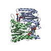
- Structure visualization
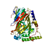
| Structure viewer | Molecule: MolmilJmol/JSmol |
|---|
- Downloads & links
-Download
| PDBx/mmCIF format | 5wvr.cif.gz | 105 KB | Display | PDBx/mmCIF format |
|---|---|---|---|---|
| PDB format | pdb5wvr.ent.gz | 77.9 KB | Display | PDB format |
| PDBx/mmJSON format | 5wvr.json.gz | Tree view | PDBx/mmJSON format | |
| Others |  Other downloads Other downloads |
-Validation report
| Arichive directory | https://data.pdbj.org/pub/pdb/validation_reports/wv/5wvrftp://data.pdbj.org/pub/pdb/validation_reports/wv/5wvr | HTTPS FTP |
|---|
-Related structure data
| Related structure data |  5h28C 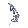 5h2aC  5h2cC  5h2dSC S: Starting model for refinement C: citing same article ( |
|---|---|
| Similar structure data |
-Links
 PDBj
PDBj


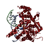
- Assembly
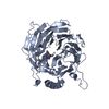
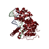
| Deposited unit | 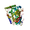
| ||||||||
|---|---|---|---|---|---|---|---|---|---|
| 1 |
| ||||||||
| Unit cell |
|
-Components
| #1: Protein | Mass: 50571.898 Da / Num. of mol.: 1 / Fragment: UNP residues 808-1240 / Mutation: 1109-1111 deletion Source method: isolated from a genetically manipulated source Source: (gene. exp.) Kluyveromyces lactis (yeast)Strain: ATCC 8585 / CBS 2359 / DSM 70799 / NBRC 1267 / NRRL Y-1140 / WM37 Gene: KLLA0_C04147g / Plasmid: modified pHIS-2 / Production host:  | ||
|---|---|---|---|
| #2: Chemical | ChemComp-CLR /   Mass: 386.654 Da / Num. of mol.: 1 / Source method: obtained synthetically / Formula: C27H46O Mass: 386.654 Da / Num. of mol.: 1 / Source method: obtained synthetically / Formula: C27H46O | ||
| #3: Chemical | ChemComp-SO4 /   Mass: 96.063 Da / Num. of mol.: 4 / Source method: obtained synthetically / Formula: SO4 Mass: 96.063 Da / Num. of mol.: 4 / Source method: obtained synthetically / Formula: SO4#4: Water | ChemComp-HOH / |  Mass: 18.015 Da / Num. of mol.: 162 / Source method: isolated from a natural source / Formula: H2O Mass: 18.015 Da / Num. of mol.: 162 / Source method: isolated from a natural source / Formula: H2O |
-Experimental details
-Experiment
| Experiment | Method: X-RAY DIFFRACTION / Number of used crystals: 1 |
|---|
- Sample preparation
| Crystal | Density Matthews: 2.06 Å3/Da / Density % sol: 40.24 % |
|---|---|
| Crystal grow | Temperature: 295 K / Method: vapor diffusion, hanging drop / pH: 9 / Details: Bicine-HCl pH 9.0, 20% PEG 3350, 0.2M MgSO4 |
-Data collection
| Diffraction | Mean temperature: 100 K |
|---|---|
| Diffraction source | Source: SYNCHROTRON / Site: PAL/PLS  / Beamline: 7A (6B, 6C1) / Wavelength: 0.9795 Å / Beamline: 7A (6B, 6C1) / Wavelength: 0.9795 Å |
| Detector | Type: ADSC QUANTUM 315 / Detector: CCD / Date: Dec 6, 2016 |
| Radiation | Monochromator: Si crystal / Protocol: SINGLE WAVELENGTH / Monochromatic (M) / Laue (L): M / Scattering type: x-ray |
| Radiation wavelength | Wavelength: 0.9795 Å / Relative weight: 1 |
| Reflection | Resolution: 2.2→50 Å / Num. obs: 20511 / % possible obs: 97.2 % / Redundancy: 3 % / Biso Wilson estimate: 26.7 Å2 / Rmerge(I) obs: 0.098 / Net I/σ(I): 23 |
| Reflection shell | Resolution: 2.2→2.24 Å / Redundancy: 3.1 % / Rmerge(I) obs: 0.373 / Mean I/σ(I) obs: 5.24 / % possible all: 97 |
- Processing
| Software |
| ||||||||||||||||||||||||||||||||||||||||||||||||||||||||
|---|---|---|---|---|---|---|---|---|---|---|---|---|---|---|---|---|---|---|---|---|---|---|---|---|---|---|---|---|---|---|---|---|---|---|---|---|---|---|---|---|---|---|---|---|---|---|---|---|---|---|---|---|---|---|---|---|---|
| Refinement | Method to determine structure: MOLECULAR REPLACEMENT Starting model: 5H2D Resolution: 2.2→22.19 Å / SU ML: 0.2 / Cross valid method: FREE R-VALUE / σ(F): 1.34 / Phase error: 23.85
| ||||||||||||||||||||||||||||||||||||||||||||||||||||||||
| Solvent computation | Shrinkage radii: 0.9 Å / VDW probe radii: 1.11 Å | ||||||||||||||||||||||||||||||||||||||||||||||||||||||||
| Refinement step | Cycle: LAST / Resolution: 2.2→22.19 Å
| ||||||||||||||||||||||||||||||||||||||||||||||||||||||||
| Refine LS restraints |
| ||||||||||||||||||||||||||||||||||||||||||||||||||||||||
| LS refinement shell |
|
