 Movie
Movie Controller

[English] 日本語
 Yorodumi
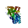
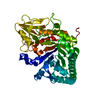
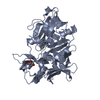
- PDB-5wvf: Crystal structure of a mutant insect group III chitinase (CAD2-E6... -
+ Open data

- Basic information
| Entry | Database: PDB / ID: 5wvf | |||||||||||||||
|---|---|---|---|---|---|---|---|---|---|---|---|---|---|---|---|---|
| Title | Crystal structure of a mutant insect group III chitinase (CAD2-E647L) from Ostrinia furnacalis | |||||||||||||||
 Components Components | Chitinase | |||||||||||||||
 Keywords Keywords | HYDROLASE / Ostrinia furnacalis / chitinase / three-dimensional structure / chitin metabolism | |||||||||||||||
| Function / homology |  Function and homology information Function and homology informationendochitinase activity / chitinase / chitin catabolic process / chitin binding / carbohydrate metabolic process / extracellular region Similarity search - Function | |||||||||||||||
| Biological species |  Ostrinia furnacalis (Asian corn borer) Ostrinia furnacalis (Asian corn borer) | |||||||||||||||
| Method |  X-RAY DIFFRACTION / SYNCHROTRON / MOLECULAR REPLACEMENT / Resolution: 2.399 Å X-RAY DIFFRACTION / SYNCHROTRON / MOLECULAR REPLACEMENT / Resolution: 2.399 Å | |||||||||||||||
 Authors Authors | Liu, T. / Zhou, Y. / Yang, Q. | |||||||||||||||
| Funding support |  China, 4items China, 4items
| |||||||||||||||
 Citation Citation | Journal: Acta Crystallogr D Struct Biol / Year: 2018 Title: The deduced role of a chitinase containing two nonsynergistic catalytic domains Authors: Liu, T. / Zhu, W. / Wang, J. / Zhou, Y. / Duan, Y. / Qu, M. / Yang, Q. | |||||||||||||||
| History |
|
- Structure visualization
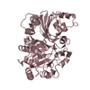
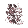
| Structure viewer | Molecule: MolmilJmol/JSmol |
|---|
- Downloads & links
-Download
| PDBx/mmCIF format | 5wvf.cif.gz | 171.6 KB | Display | PDBx/mmCIF format |
|---|---|---|---|---|
| PDB format | pdb5wvf.ent.gz | 134 KB | Display | PDB format |
| PDBx/mmJSON format | 5wvf.json.gz | Tree view | PDBx/mmJSON format | |
| Others |  Other downloads Other downloads |
-Validation report
| Arichive directory | https://data.pdbj.org/pub/pdb/validation_reports/wv/5wvfftp://data.pdbj.org/pub/pdb/validation_reports/wv/5wvf | HTTPS FTP |
|---|
-Related structure data
| Related structure data | 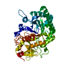 5wupC  5wusSC 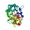 5wv8C 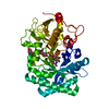 5wv9C 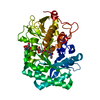 5wvbC 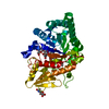 5wvgC 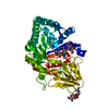 5wvhC C: citing same article ( S: Starting model for refinement |
|---|---|
| Similar structure data |


-Links
 PDBj
PDBj- Assembly
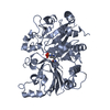
| Deposited unit | 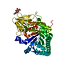
| |||||||||
|---|---|---|---|---|---|---|---|---|---|---|
| 1 |
| |||||||||
| Unit cell |
| |||||||||
| Components on special symmetry positions |
|
-Components
| #1: Protein | Mass: 52409.617 Da / Num. of mol.: 1 / Fragment: UNP residues 528-987 / Mutation: E647L Source method: isolated from a genetically manipulated source Source: (gene. exp.) Ostrinia furnacalis (Asian corn borer) / Production host:  Komagataella pastoris CBS 7435 (fungus) / References: UniProt: U5LUV7, chitinase Komagataella pastoris CBS 7435 (fungus) / References: UniProt: U5LUV7, chitinase |
|---|---|
| #2: Sugar | ChemComp-NAG /   Type: D-saccharide, beta linking / Mass: 221.208 Da / Num. of mol.: 1 Type: D-saccharide, beta linking / Mass: 221.208 Da / Num. of mol.: 1Source method: isolated from a genetically manipulated source Formula: C8H15NO6 |
| #3: Water | ChemComp-HOH /  Mass: 18.015 Da / Num. of mol.: 101 / Source method: isolated from a natural source / Formula: H2O Mass: 18.015 Da / Num. of mol.: 101 / Source method: isolated from a natural source / Formula: H2O |
| Has protein modification | Y |
-Experimental details
-Experiment
| Experiment | Method: X-RAY DIFFRACTION / Number of used crystals: 1 |
|---|
- Sample preparation
| Crystal | Density Matthews: 2.14 Å3/Da / Density % sol: 42.64 % |
|---|---|
| Crystal grow | Temperature: 277 K / Method: vapor diffusion, hanging drop / pH: 8.1 / Details: 200mM tri-sodium citrate dihydrate, 20% PEG3350 |
-Data collection
| Diffraction | Mean temperature: 100 K |
|---|---|
| Diffraction source | Source: SYNCHROTRON / Site: SSRF / Beamline: BL17U / Wavelength: 0.97915 Å |
| Detector | Type: ADSC QUANTUM 315r / Detector: CCD / Date: Jun 15, 2015 |
| Radiation | Protocol: SINGLE WAVELENGTH / Monochromatic (M) / Laue (L): M / Scattering type: x-ray |
| Radiation wavelength | Wavelength: 0.97915 Å / Relative weight: 1 |
| Reflection | Resolution: 2.399→30 Å / Num. obs: 18731 / % possible obs: 99.6 % / Redundancy: 13.5 % / Rsym value: 0.067 / Net I/σ(I): 11.1 |
| Reflection shell | Resolution: 2.399→2.44 Å / Redundancy: 13.9 % / Rsym value: 0.381 / % possible all: 100 |
- Processing
| Software |
| ||||||||||||||||||||||||||||||||||||||||||||||||||||||||||||||||||||||||||||||||||||||||||||||||||
|---|---|---|---|---|---|---|---|---|---|---|---|---|---|---|---|---|---|---|---|---|---|---|---|---|---|---|---|---|---|---|---|---|---|---|---|---|---|---|---|---|---|---|---|---|---|---|---|---|---|---|---|---|---|---|---|---|---|---|---|---|---|---|---|---|---|---|---|---|---|---|---|---|---|---|---|---|---|---|---|---|---|---|---|---|---|---|---|---|---|---|---|---|---|---|---|---|---|---|---|
| Refinement | Method to determine structure: MOLECULAR REPLACEMENT Starting model: 5wus Resolution: 2.399→28.883 Å / SU ML: 0.24 / Cross valid method: FREE R-VALUE / σ(F): 1.34 / Phase error: 22.77 / Stereochemistry target values: ML
| ||||||||||||||||||||||||||||||||||||||||||||||||||||||||||||||||||||||||||||||||||||||||||||||||||
| Solvent computation | Shrinkage radii: 0.9 Å / VDW probe radii: 1.11 Å / Solvent model: FLAT BULK SOLVENT MODEL | ||||||||||||||||||||||||||||||||||||||||||||||||||||||||||||||||||||||||||||||||||||||||||||||||||
| Refinement step | Cycle: LAST / Resolution: 2.399→28.883 Å
| ||||||||||||||||||||||||||||||||||||||||||||||||||||||||||||||||||||||||||||||||||||||||||||||||||
| Refine LS restraints |
| ||||||||||||||||||||||||||||||||||||||||||||||||||||||||||||||||||||||||||||||||||||||||||||||||||
| LS refinement shell |
| ||||||||||||||||||||||||||||||||||||||||||||||||||||||||||||||||||||||||||||||||||||||||||||||||||
| Refinement TLS params. | Method: refined / Origin x: -9.2492 Å / Origin y: 17.2832 Å / Origin z: 18.2217 Å
| ||||||||||||||||||||||||||||||||||||||||||||||||||||||||||||||||||||||||||||||||||||||||||||||||||
| Refinement TLS group | Selection details: all |
