 Movie
Movie Controller


+ Open data

- Basic information
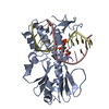
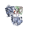
| Entry | Database: PDB / ID: 5wa3 | ||||||
|---|---|---|---|---|---|---|---|
| Title | Pyridine synthase, TbtD, from thiomuracin biosynthesis | ||||||
 Components Components | Pyridine synthase TbtD | ||||||
 Keywords Keywords | BIOSYNTHETIC PROTEIN / pyridine synthase / Diels-Alder / thiopeptide / thiomuracin | ||||||
| Function / homology | : / Thiopeptide-type bacteriocin biosynthesis domain / Lantibiotic biosynthesis dehydratase C-term / Thiopeptide-type bacteriocin biosynthesis domain-containing protein Function and homology information Function and homology information | ||||||
| Biological species |  Thermobispora bispora (bacteria) Thermobispora bispora (bacteria) | ||||||
| Method |  X-RAY DIFFRACTION / SYNCHROTRON / MOLECULAR REPLACEMENT / Resolution: 2.8 Å X-RAY DIFFRACTION / SYNCHROTRON / MOLECULAR REPLACEMENT / Resolution: 2.8 Å | ||||||
 Authors Authors | Cogan, D.P. / Nair, S.K. | ||||||
 Citation Citation | Journal: Proc. Natl. Acad. Sci. U.S.A. / Year: 2017 Title: Structural insights into enzymatic [4+2] aza-cycloaddition in thiopeptide antibiotic biosynthesis. Authors: Cogan, D.P. / Hudson, G.A. / Zhang, Z. / Pogorelov, T.V. / van der Donk, W.A. / Mitchell, D.A. / Nair, S.K. | ||||||
| History |
|
- Structure visualization
| Structure viewer | Molecule: MolmilJmol/JSmol |
|---|
- Downloads & links
-Download
| PDBx/mmCIF format | 5wa3.cif.gz | 133 KB | Display | PDBx/mmCIF format |
|---|---|---|---|---|
| PDB format | pdb5wa3.ent.gz | 104.3 KB | Display | PDB format |
| PDBx/mmJSON format | 5wa3.json.gz | Tree view | PDBx/mmJSON format | |
| Others |  Other downloads Other downloads |
-Validation report
| Arichive directory | https://data.pdbj.org/pub/pdb/validation_reports/wa/5wa3ftp://data.pdbj.org/pub/pdb/validation_reports/wa/5wa3 | HTTPS FTP |
|---|
-Related structure data
| Related structure data |  5w98SC 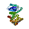 5w99C 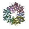 5wa4C S: Starting model for refinement C: citing same article ( |
|---|---|
| Similar structure data |


-Links
 PDBj
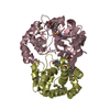
PDBj- Assembly
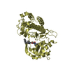
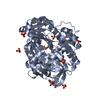
| Deposited unit | 
| ||||||||
|---|---|---|---|---|---|---|---|---|---|
| 1 | 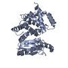
| ||||||||
| 2 | 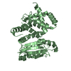
| ||||||||
| 3 | 
| ||||||||
| Unit cell |
|
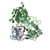
-Components
| #1: Protein | Mass: 40320.555 Da / Num. of mol.: 2 Source method: isolated from a genetically manipulated source Source: (gene. exp.) Thermobispora bispora (bacteria)Strain: ATCC 19993 / DSM 43833 / CBS 139.67 / JCM 10125 / NBRC 14880 / R51 Gene: Tbis_0552 / Plasmid: pRARE / Production host: Sequence details | TbtD in the NCBI database: WP_050760415.1 | |
|---|
-Experimental details
-Experiment
| Experiment | Method: X-RAY DIFFRACTION / Number of used crystals: 1 |
|---|
- Sample preparation
| Crystal | Density Matthews: 4.79 Å3/Da / Density % sol: 74.31 % |
|---|---|
| Crystal grow | Temperature: 289 K / Method: vapor diffusion, hanging drop / pH: 6.5 / Details: 0.4 M NaH2PO4, 1 M KH2PO4 pH 6.5 |
-Data collection
| Diffraction | Mean temperature: 100 K |
|---|---|
| Diffraction source | Source: SYNCHROTRON / Site: APS  / Beamline: 21-ID-G / Wavelength: 0.97857 Å / Beamline: 21-ID-G / Wavelength: 0.97857 Å |
| Detector | Type: MARMOSAIC 300 mm CCD / Detector: CCD / Date: Jun 13, 2015 |
| Radiation | Protocol: SINGLE WAVELENGTH / Monochromatic (M) / Laue (L): M / Scattering type: x-ray |
| Radiation wavelength | Wavelength: 0.97857 Å / Relative weight: 1 |
| Reflection | Resolution: 2.8→55.08 Å / Num. obs: 34569 / % possible obs: 93.9 % / Redundancy: 6.3 % / Rmerge(I) obs: 0.117 / Net I/σ(I): 10 |
- Processing
| Software |
| ||||||||||||||||||||||||||||||||||||||||||||||||||||||||||||||||||||||||||||||||||||||||||||||||||||||||||||||||||||||||||||||||||||||||||||||||||||||||||||||||||||||||||||||||||||||
|---|---|---|---|---|---|---|---|---|---|---|---|---|---|---|---|---|---|---|---|---|---|---|---|---|---|---|---|---|---|---|---|---|---|---|---|---|---|---|---|---|---|---|---|---|---|---|---|---|---|---|---|---|---|---|---|---|---|---|---|---|---|---|---|---|---|---|---|---|---|---|---|---|---|---|---|---|---|---|---|---|---|---|---|---|---|---|---|---|---|---|---|---|---|---|---|---|---|---|---|---|---|---|---|---|---|---|---|---|---|---|---|---|---|---|---|---|---|---|---|---|---|---|---|---|---|---|---|---|---|---|---|---|---|---|---|---|---|---|---|---|---|---|---|---|---|---|---|---|---|---|---|---|---|---|---|---|---|---|---|---|---|---|---|---|---|---|---|---|---|---|---|---|---|---|---|---|---|---|---|---|---|---|---|
| Refinement | Method to determine structure: MOLECULAR REPLACEMENT Starting model: 5W98 Resolution: 2.8→104.52 Å / Cor.coef. Fo:Fc: 0.947 / Cor.coef. Fo:Fc free: 0.927 / SU B: 11.451 / SU ML: 0.221 / Cross valid method: THROUGHOUT / ESU R: 0.347 / ESU R Free: 0.271 / Stereochemistry target values: MAXIMUM LIKELIHOOD / Details: HYDROGENS HAVE BEEN ADDED IN THE RIDING POSITIONS
| ||||||||||||||||||||||||||||||||||||||||||||||||||||||||||||||||||||||||||||||||||||||||||||||||||||||||||||||||||||||||||||||||||||||||||||||||||||||||||||||||||||||||||||||||||||||
| Solvent computation | Ion probe radii: 0.8 Å / Shrinkage radii: 0.8 Å / VDW probe radii: 1.2 Å / Solvent model: MASK | ||||||||||||||||||||||||||||||||||||||||||||||||||||||||||||||||||||||||||||||||||||||||||||||||||||||||||||||||||||||||||||||||||||||||||||||||||||||||||||||||||||||||||||||||||||||
| Displacement parameters | Biso mean: 88.879 Å2
| ||||||||||||||||||||||||||||||||||||||||||||||||||||||||||||||||||||||||||||||||||||||||||||||||||||||||||||||||||||||||||||||||||||||||||||||||||||||||||||||||||||||||||||||||||||||
| Refinement step | Cycle: 1 / Resolution: 2.8→104.52 Å
| ||||||||||||||||||||||||||||||||||||||||||||||||||||||||||||||||||||||||||||||||||||||||||||||||||||||||||||||||||||||||||||||||||||||||||||||||||||||||||||||||||||||||||||||||||||||
| Refine LS restraints |
|