 Movie
Movie Controller

[English] 日本語
 Yorodumi
- PDB-5w8i: Crystal Structure of Lactate Dehydrogenase A in complex with inhi... -
+ Open data

- Basic information
| Entry | Database: PDB / ID: 5w8i | ||||||
|---|---|---|---|---|---|---|---|
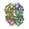
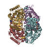
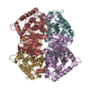
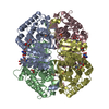
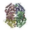
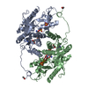
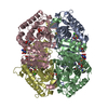
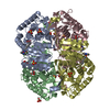
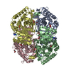
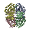
| Title | Crystal Structure of Lactate Dehydrogenase A in complex with inhibitor compound 23 and Zinc | ||||||
 Components Components | L-lactate dehydrogenase A chain | ||||||
 Keywords Keywords | OXIDOREDUCTASE / oxidoreductase inhibitor | ||||||
| Function / homology |  Function and homology information Function and homology informationsperm fibrous sheath / oxidoreductase complex / pyruvate catabolic process / L-lactate dehydrogenase / L-lactate dehydrogenase (NAD+) activity / Pyruvate metabolism / lactate metabolic process / : / substantia nigra development / Regulation of pyruvate metabolism ...sperm fibrous sheath / oxidoreductase complex / pyruvate catabolic process / L-lactate dehydrogenase / L-lactate dehydrogenase (NAD+) activity / Pyruvate metabolism / lactate metabolic process / : / substantia nigra development / Regulation of pyruvate metabolism / glycolytic process / cadherin binding / mitochondrion / extracellular exosome / membrane / identical protein binding / nucleus / cytosol Similarity search - Function | ||||||
| Biological species |  Homo sapiens (human) Homo sapiens (human) | ||||||
| Method |  X-RAY DIFFRACTION / Resolution: 1.95 Å X-RAY DIFFRACTION / Resolution: 1.95 Å | ||||||
 Authors Authors | Lukacs, C.M. / Abendroth, J. | ||||||
 Citation Citation | Journal: J. Med. Chem. / Year: 2017 Title: Discovery and Optimization of Potent, Cell-Active Pyrazole-Based Inhibitors of Lactate Dehydrogenase (LDH). Authors: Rai, G. / Brimacombe, K.R. / Mott, B.T. / Urban, D.J. / Hu, X. / Yang, S.M. / Lee, T.D. / Cheff, D.M. / Kouznetsova, J. / Benavides, G.A. / Pohida, K. / Kuenstner, E.J. / Luci, D.K. / ...Authors: Rai, G. / Brimacombe, K.R. / Mott, B.T. / Urban, D.J. / Hu, X. / Yang, S.M. / Lee, T.D. / Cheff, D.M. / Kouznetsova, J. / Benavides, G.A. / Pohida, K. / Kuenstner, E.J. / Luci, D.K. / Lukacs, C.M. / Davies, D.R. / Dranow, D.M. / Zhu, H. / Sulikowski, G. / Moore, W.J. / Stott, G.M. / Flint, A.J. / Hall, M.D. / Darley-Usmar, V.M. / Neckers, L.M. / Dang, C.V. / Waterson, A.G. / Simeonov, A. / Jadhav, A. / Maloney, D.J. | ||||||
| History |
|
- Structure visualization
| Structure viewer | Molecule: MolmilJmol/JSmol |
|---|
- Downloads & links
-Download
| PDBx/mmCIF format | 5w8i.cif.gz | 513.5 KB | Display | PDBx/mmCIF format |
|---|---|---|---|---|
| PDB format | pdb5w8i.ent.gz | 420.7 KB | Display | PDB format |
| PDBx/mmJSON format | 5w8i.json.gz | Tree view | PDBx/mmJSON format | |
| Others |  Other downloads Other downloads |
-Validation report
| Arichive directory | https://data.pdbj.org/pub/pdb/validation_reports/w8/5w8iftp://data.pdbj.org/pub/pdb/validation_reports/w8/5w8i | HTTPS FTP |
|---|
-Related structure data
| Related structure data |  5w8hC  5w8jC  5w8kC  5w8lC  4jnkS S: Starting model for refinement C: citing same article ( |
|---|---|
| Similar structure data |
-Links
 PDBj
PDBj


- Assembly
| Deposited unit | 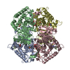
| ||||||||
|---|---|---|---|---|---|---|---|---|---|
| 1 |
| ||||||||
| Unit cell |
|
-Components
-Protein , 1 types, 4 molecules ABCD
| #1: Protein | Mass: 36734.672 Da / Num. of mol.: 4 Source method: isolated from a genetically manipulated source Source: (gene. exp.) Homo sapiens (human) / Gene: LDHA, PIG19 / Production host:  |
|---|
-Non-polymers , 7 types, 930 molecules 


| #2: Chemical | ChemComp-ZN /  Mass: 65.409 Da / Num. of mol.: 5 / Source method: obtained synthetically / Formula: Zn Mass: 65.409 Da / Num. of mol.: 5 / Source method: obtained synthetically / Formula: Zn#3: Chemical |  Mass: 323.275 Da / Num. of mol.: 3 / Source method: obtained synthetically / Formula: C13H7F2N3O3S Mass: 323.275 Da / Num. of mol.: 3 / Source method: obtained synthetically / Formula: C13H7F2N3O3S#4: Chemical | ChemComp-CIT /  Mass: 192.124 Da / Num. of mol.: 4 / Source method: obtained synthetically / Formula: C6H8O7 Mass: 192.124 Da / Num. of mol.: 4 / Source method: obtained synthetically / Formula: C6H8O7#5: Chemical | ChemComp-DMS /  Mass: 78.133 Da / Num. of mol.: 4 / Source method: obtained synthetically / Formula: C2H6OS / Comment: DMSO, precipitant*YM Mass: 78.133 Da / Num. of mol.: 4 / Source method: obtained synthetically / Formula: C2H6OS / Comment: DMSO, precipitant*YM#6: Chemical | ChemComp-MLA / |  Mass: 104.061 Da / Num. of mol.: 1 / Source method: obtained synthetically / Formula: C3H4O4 Mass: 104.061 Da / Num. of mol.: 1 / Source method: obtained synthetically / Formula: C3H4O4#7: Chemical | ChemComp-GOL / |  Mass: 92.094 Da / Num. of mol.: 1 / Source method: obtained synthetically / Formula: C3H8O3 Mass: 92.094 Da / Num. of mol.: 1 / Source method: obtained synthetically / Formula: C3H8O3#8: Water | ChemComp-HOH / | Mass: 18.015 Da / Num. of mol.: 912 / Source method: isolated from a natural source / Formula: H2O |
|---|
-Experimental details
-Experiment
| Experiment | Method: X-RAY DIFFRACTION / Number of used crystals: 1 |
|---|
- Sample preparation
| Crystal | Density Matthews: 2.34 Å3/Da / Density % sol: 47 % |
|---|---|
| Crystal grow | Temperature: 289 K / Method: vapor diffusion / pH: 6.8 Details: 24% PEG 3350, 200 mM Sodium Malonate, pH 6.8, 10% Glycerol |
-Data collection
| Diffraction | Mean temperature: 100 K |
|---|---|
| Diffraction source | Source: ROTATING ANODE / Type: RIGAKU FR-E+ SUPERBRIGHT / Wavelength: 1.54178 Å |
| Detector | Type: RIGAKU SATURN 944+ / Detector: CCD / Date: Jan 27, 2014 |
| Radiation | Protocol: SINGLE WAVELENGTH / Monochromatic (M) / Laue (L): M / Scattering type: x-ray |
| Radiation wavelength | Wavelength: 1.54178 Å / Relative weight: 1 |
| Reflection | Resolution: 1.95→50 Å / Num. obs: 102092 / % possible obs: 99.7 % / Observed criterion σ(I): -3 / Redundancy: 9.4 % / Biso Wilson estimate: 26.99 Å2 / Rmerge(I) obs: 0.083 / Net I/σ(I): 21.84 |
| Reflection shell | Resolution: 1.95→2 Å / Redundancy: 5.1 % / Rmerge(I) obs: 0.556 / Mean I/σ(I) obs: 3.2 / % possible all: 99.6 |
- Processing
| Software |
| |||||||||||||||||||||||||||||||||||||||||||||||||||||||||||||||||||||||||||||||||||||||||||||||||||||||||||||||||||||||||||||
|---|---|---|---|---|---|---|---|---|---|---|---|---|---|---|---|---|---|---|---|---|---|---|---|---|---|---|---|---|---|---|---|---|---|---|---|---|---|---|---|---|---|---|---|---|---|---|---|---|---|---|---|---|---|---|---|---|---|---|---|---|---|---|---|---|---|---|---|---|---|---|---|---|---|---|---|---|---|---|---|---|---|---|---|---|---|---|---|---|---|---|---|---|---|---|---|---|---|---|---|---|---|---|---|---|---|---|---|---|---|---|---|---|---|---|---|---|---|---|---|---|---|---|---|---|---|---|
| Refinement | Starting model: IN-HOUSE APO SOLVED WITH MODEL 4JNK Resolution: 1.95→50 Å / Cor.coef. Fo:Fc: 0.967 / Cor.coef. Fo:Fc free: 0.948 / SU B: 5.974 / SU ML: 0.086 / Cross valid method: THROUGHOUT / σ(F): 0 / ESU R: 0.136 / ESU R Free: 0.126 Details: HYDROGENS HAVE BEEN ADDED IN THE RIDING POSITIONS U VALUES : WITH TLS ADDED
| |||||||||||||||||||||||||||||||||||||||||||||||||||||||||||||||||||||||||||||||||||||||||||||||||||||||||||||||||||||||||||||
| Solvent computation | Ion probe radii: 0.8 Å / Shrinkage radii: 0.8 Å / VDW probe radii: 1.2 Å | |||||||||||||||||||||||||||||||||||||||||||||||||||||||||||||||||||||||||||||||||||||||||||||||||||||||||||||||||||||||||||||
| Displacement parameters | Biso max: 68.62 Å2 / Biso mean: 21.13 Å2 / Biso min: 9.45 Å2
| |||||||||||||||||||||||||||||||||||||||||||||||||||||||||||||||||||||||||||||||||||||||||||||||||||||||||||||||||||||||||||||
| Refinement step | Cycle: final / Resolution: 1.95→50 Å
| |||||||||||||||||||||||||||||||||||||||||||||||||||||||||||||||||||||||||||||||||||||||||||||||||||||||||||||||||||||||||||||
| LS refinement shell | Resolution: 1.95→2 Å / Rfactor Rfree error: 0 / Total num. of bins used: 20
| |||||||||||||||||||||||||||||||||||||||||||||||||||||||||||||||||||||||||||||||||||||||||||||||||||||||||||||||||||||||||||||
| Refinement TLS params. | Method: refined / Refine-ID: X-RAY DIFFRACTION
| |||||||||||||||||||||||||||||||||||||||||||||||||||||||||||||||||||||||||||||||||||||||||||||||||||||||||||||||||||||||||||||
| Refinement TLS group |
|
