[English] 日本語
Yorodumi
- PDB-5w3n: Molecular structure of FUS low sequence complexity domain protein... -

+
Open data


ID or keywords:

Loading...

-
Basic information

Entry
Database: PDB / ID: 5w3n
TitleMolecular structure of FUS low sequence complexity domain protein fibrils
ComponentsRNA-binding protein FUS
KeywordsPROTEIN FIBRIL / Low complexity sequence domain / RNA transport / RNA Granule / intrinsically disordered protein
Function / homology
Function and homology information


mRNA stabilization / positive regulation of double-strand break repair via homologous recombination / intracellular non-membrane-bounded organelle / regulation of RNA splicing / Processing of Capped Intron-Containing Pre-mRNA / molecular condensate scaffold activity / mRNA Splicing - Major Pathway / RNA splicing / mRNA 3'-UTR binding / transcription coregulator activity ...mRNA stabilization / positive regulation of double-strand break repair via homologous recombination / intracellular non-membrane-bounded organelle / regulation of RNA splicing / Processing of Capped Intron-Containing Pre-mRNA / molecular condensate scaffold activity / mRNA Splicing - Major Pathway / RNA splicing / mRNA 3'-UTR binding / transcription coregulator activity / protein homooligomerization / amyloid fibril formation / transcription coactivator activity / chromatin binding / regulation of DNA-templated transcription / regulation of transcription by RNA polymerase II / DNA binding / RNA binding / nucleoplasm / identical protein binding / metal ion binding / nucleus / cytoplasm
Similarity search - Function
TAF15/EWS/TLS family / Zinc finger domain / Zn-finger in Ran binding protein and others / Zinc finger RanBP2 type profile. / Zinc finger RanBP2-type signature. / Zinc finger, RanBP2-type superfamily / Zinc finger, RanBP2-type / RNA recognition motif / RNA recognition motif / Eukaryotic RNA Recognition Motif (RRM) profile. ...TAF15/EWS/TLS family / Zinc finger domain / Zn-finger in Ran binding protein and others / Zinc finger RanBP2 type profile. / Zinc finger RanBP2-type signature. / Zinc finger, RanBP2-type superfamily / Zinc finger, RanBP2-type / RNA recognition motif / RNA recognition motif / Eukaryotic RNA Recognition Motif (RRM) profile. / RNA recognition motif domain / RNA-binding domain superfamily / Nucleotide-binding alpha-beta plait domain superfamily
Similarity search - Domain/homology
RNA-binding protein FUS
Similarity search - Component
Biological speciesHomo sapiens (human)
MethodSOLID-STATE NMR / simulated annealing
AuthorsMurray, D.T. / Kato, M. / Lin, Y. / Thurber, K. / Hung, I. / McKnight, S. / Tycko, R.
Funding support United States, 2items
OrganizationGrant numberCountry
National Institutes of Health/National Institute of General Medical Sciences (NIH/NIGMS)5U01GM107623-03 United States
National Institutes of Health/National Institute of General Medical Sciences (NIH/NIGMS)1FI2GM117604-01 United States
CitationJournal: Cell / Year: 2017
Title: Structure of FUS Protein Fibrils and Its Relevance to Self-Assembly and Phase Separation of Low-Complexity Domains.
Authors: Murray, D.T. / Kato, M. / Lin, Y. / Thurber, K.R. / Hung, I. / McKnight, S.L. / Tycko, R.
History
DepositionJun 8, 2017Deposition site: RCSB / Processing site: RCSB
Revision 1.0Sep 27, 2017Provider: repository / Type: Initial release
Revision 1.1Oct 18, 2017Group: Author supporting evidence / Category: pdbx_audit_support / Item: _pdbx_audit_support.funding_organization
Revision 1.2Apr 10, 2019Group: Data collection / Database references / Category: citation / citation_author / pdbx_nmr_software
Item: _citation.journal_abbrev / _citation.journal_id_CSD ..._citation.journal_abbrev / _citation.journal_id_CSD / _citation.journal_id_ISSN / _citation.journal_volume / _citation.page_first / _citation.page_last / _citation.pdbx_database_id_DOI / _citation.pdbx_database_id_PubMed / _citation.title / _citation.year / _citation_author.name / _pdbx_nmr_software.name
Revision 1.3Jan 1, 2020Group: Author supporting evidence / Data collection / Category: pdbx_audit_support / pdbx_nmr_spectrometer
Item: _pdbx_audit_support.funding_organization / _pdbx_nmr_spectrometer.model
Revision 1.4Jun 14, 2023Group: Database references / Other / Category: database_2 / pdbx_database_status
Item: _database_2.pdbx_DOI / _database_2.pdbx_database_accession / _pdbx_database_status.status_code_nmr_data
Revision 1.5May 15, 2024Group: Data collection / Database references / Category: chem_comp_atom / chem_comp_bond / database_2 / Item: _database_2.pdbx_DOI

-
Structure visualization

Structure viewerMolecule:
MolmilJmol/JSmol

Downloads & links

-
Assembly

Deposited unit
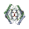
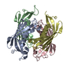
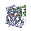
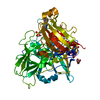
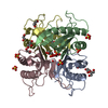
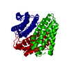
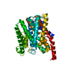
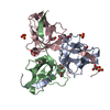
A: RNA-binding protein FUS
B: RNA-binding protein FUS
C: RNA-binding protein FUS
D: RNA-binding protein FUS
E: RNA-binding protein FUS
F: RNA-binding protein FUS
G: RNA-binding protein FUS
H: RNA-binding protein FUS
I: RNA-binding protein FUS


Theoretical massNumber of molelcules
Total (without water)223,8299
Polymers223,8299
Non-polymers00
Water0
1


  • Idetical with deposited unit
  • defined by author&software
  • Evidence: microscopy, Mass-per-length measurements were performed on a transmission electron microscope to determine the repeating unit in the protein fibril. Atomic force microscopy images support ...Evidence: microscopy, Mass-per-length measurements were performed on a transmission electron microscope to determine the repeating unit in the protein fibril. Atomic force microscopy images support the diameter of the calculated macromolecular assembly.
TypeNameSymmetry operationNumber
identity operation1_5551
Buried area35890 Å2
ΔGint-48 kcal/mol
Surface area17380 Å2
MethodPISA
NMR ensembles
DataCriteria
Number of conformers (submitted / calculated)20 / 4928structures with no violations, lowest energy, and derived from one of 44 independent calculations
RepresentativeModel #1lowest energy

-
Components

#1: Protein
RNA-binding protein FUS / / 75 kDa DNA-pairing protein / Oncogene FUS / Oncogene TLS / POMp75 / Translocated in liposarcoma protein


Mass: 24869.846 Da / Num. of mol.: 9 / Fragment: UNP residues 2-214
Source method: isolated from a genetically manipulated source
Source: (gene. exp.) Homo sapiens (human) / Gene: FUS, TLS
Production host: Escherichia coli 'BL21-Gold(DE3)pLysS AG' (bacteria)
References: UniProt: P35637

-
Experimental details

-
Experiment

ExperimentMethod: SOLID-STATE NMR
NMR experiment
Conditions-IDExperiment-IDSolution-IDSample stateSpectrometer-IDType
111isotropic13D NCACX
121isotropic13D NCOCX
131isotropic33D CONCA
141isotropic33D CANCX
154isotropic413C-13C PITHIRDS-CT
165isotropic413C-13C PITHIRDS-CT
171isotropic215N-15N BARE
182isotropic215N-15N BARE
192isotropic213C-13C DARR
1103isotropic213C-13C DARR
1112isotropic13D NCACX
1123isotropic13D NCOCX

-
Sample preparation

Details
TypeSolution-IDContentsDetailsLabelSolvent system
solid10.28 mg/uL [U-99% 13C; U-99% 15N] FUS-LC Fibril, 100% H2OUniformly 15N,13C labeled FUS low sequence complexity domain protein fibrilsU-FUS-LC100% H2O
solid20.28 mg/uL 99% 15N, 99% 2-13C Glycerol FUS-LC Fibril, 100% H2OUniformly 15N, 2-13C glycerol labeled FUS low sequence complexity domain protein fibrils2-Glc-FUS-LC100% H2O
solid30.28 mg/uL 99% 15N, 99% 1,3-13C Glycerol FUS-LC Fibril, 100% H2OUniformly 15N, 1,3-13C glycerol labeled FUS low sequence complexity domain protein fibrils1,3-Glc-FUS-LC100% H2O
solid40.23 mg/uL 99% 1-13C Tyrosine FUS-LC Fibril, 100% H2O1-13C Tyrosine labeled FUS low sequence complexity domain fibrils1-13C-Tyr-FUS-LC100% H2O
solid50.23 mg/uL 99% 1-13C Threonine FUS-LC Fibril, 100% H2O1-13C Threonine labeled FUS low sequence complexity domain fibrils1-13C-Thr-FUS-LC100% H2O
Sample
Conc. (mg/ml)ComponentIsotopic labelingSolution-ID
0.28 mg/uLFUS-LC Fibril[U-99% 13C; U-99% 15N]1
0.28 mg/uLFUS-LC Fibril99% 15N, 99% 2-13C Glycerol2
0.28 mg/uLFUS-LC Fibril99% 15N, 99% 1,3-13C Glycerol3
0.23 mg/uLFUS-LC Fibril99% 1-13C Tyrosine4
0.23 mg/uLFUS-LC Fibril99% 1-13C Threonine5
Sample conditionsDetails: Samples are hydrated protein fibrils in 20 mM sodium phosphate, pH 7.4
Ionic strength: 20 mM / Label: conditions_1 / pH: 7.4 / Pressure: 1 atm / Temperature: 298 K

-
NMR measurement

NMR spectrometer
TypeManufacturerModelField strength (MHz)Spectrometer-ID
Bruker AVANCE IIIBrukerAVANCE III895.11
Varian InfinityVarianInfinity746.12
Varian InfinityPlusVarianInfinityPlus599.23
Bruker AVANCE IIIBrukerAVANCE III400.64

-
Processing

NMR software
NameVersionDeveloperClassification
MCASSIGN2BKan-Nian Hu, Wei Qiang, and Robert Tyckochemical shift assignment
NMRPipe8.9Delaglio, Grzesiek, Vuister, Zhu, Pfeifer and Baxprocessing
SparkyGoddarddata analysis
X-PLOR NIH2.45Schwieters, Kuszewski, Tjandra and Clorerefinement
RefinementMethod: simulated annealing / Software ordinal: 4
Details: Calculate 448 initial folds for an extended FUS monomer with nine identical copies. Select 10% lowest energy for further calculations. Calculate 112 structures for each lowest energy model ...Details: Calculate 448 initial folds for an extended FUS monomer with nine identical copies. Select 10% lowest energy for further calculations. Calculate 112 structures for each lowest energy model in first round of calculations. Analyze 10% lowest energy structures in each run and select based on stated selection criterion.
NMR representativeSelection criteria: lowest energy
NMR ensembleConformer selection criteria: structures with no violations, lowest energy, and derived from one of 44 independent calculations
Conformers calculated total number: 4928 / Conformers submitted total number: 20

+
About Yorodumi

-
News

-
Feb 9, 2022. New format data for meta-information of EMDB entries

New format data for meta-information of EMDB entries

  • Version 3 of the EMDB header file is now the official format.
  • The previous official version 1.9 will be removed from the archive.

Related info.:EMDB header

External links:wwPDB to switch to version 3 of the EMDB data model

-
Aug 12, 2020. Covid-19 info

Covid-19 info

URL: https://pdbj.org/emnavi/covid19.php

New page: Covid-19 featured information page in EM Navigator.

Related info.:Covid-19 info / Mar 5, 2020. Novel coronavirus structure data

+
Mar 5, 2020. Novel coronavirus structure data

Novel coronavirus structure data

Related info.:Yorodumi Speices / Aug 12, 2020. Covid-19 info

External links:COVID-19 featured content - PDBj / Molecule of the Month (242):Coronavirus Proteases

+
Jan 31, 2019. EMDB accession codes are about to change! (news from PDBe EMDB page)

EMDB accession codes are about to change! (news from PDBe EMDB page)

  • The allocation of 4 digits for EMDB accession codes will soon come to an end. Whilst these codes will remain in use, new EMDB accession codes will include an additional digit and will expand incrementally as the available range of codes is exhausted. The current 4-digit format prefixed with “EMD-” (i.e. EMD-XXXX) will advance to a 5-digit format (i.e. EMD-XXXXX), and so on. It is currently estimated that the 4-digit codes will be depleted around Spring 2019, at which point the 5-digit format will come into force.
  • The EM Navigator/Yorodumi systems omit the EMD- prefix.

Related info.:Q: What is EMD? / ID/Accession-code notation in Yorodumi/EM Navigator

External links:EMDB Accession Codes are Changing Soon! / Contact to PDBj

+
Jul 12, 2017. Major update of PDB

Major update of PDB

  • wwPDB released updated PDB data conforming to the new PDBx/mmCIF dictionary.
  • This is a major update changing the version number from 4 to 5, and with Remediation, in which all the entries are updated.
  • In this update, many items about electron microscopy experimental information are reorganized (e.g. em_software).
  • Now, EM Navigator and Yorodumi are based on the updated data.

External links:wwPDB Remediation / Enriched Model Files Conforming to OneDep Data Standards Now Available in the PDB FTP Archive

-
Yorodumi

Thousand views of thousand structures

  • Yorodumi is a browser for structure data from EMDB, PDB, SASBDB, etc.
  • This page is also the successor to EM Navigator detail page, and also detail information page/front-end page for Omokage search.
  • The word "yorodu" (or yorozu) is an old Japanese word meaning "ten thousand". "mi" (miru) is to see.

Related info.:EMDB / PDB / SASBDB / Comparison of 3 databanks / Yorodumi Search / Aug 31, 2016. New EM Navigator & Yorodumi / Yorodumi Papers / Jmol/JSmol / Function and homology information / Changes in new EM Navigator and Yorodumi

Read more