[English] 日本語
Yorodumi
- PDB-3gi7: Crystal structure of a duf1311 family protein (pp0307) from pseud... -

+
Open data


ID or keywords:

Loading...

-
Basic information

Entry
Database: PDB / ID: 3gi7
TitleCrystal structure of a duf1311 family protein (pp0307) from pseudomonas putida kt2440 at 1.85 A resolution
ComponentsSecreted protein of unknown function DUF1311
KeywordsUNKNOWN FUNCTION / Secreted protein / structural genomics / Joint Center for Structural Genomics / JCSG / Protein Structure Initiative / PSI-2
Function / homologySubstrate Binding Domain Of Dnak; Chain:A; Domain 2 - #180 / Lysozyme inhibitor LprI, N-terminal / Lysozyme inhibitor LprI / Substrate Binding Domain Of Dnak; Chain:A; Domain 2 / Up-down Bundle / Mainly Alpha / Unknown ligand / Lysozyme inhibitor LprI-like N-terminal domain-containing protein
Function and homology information
Biological speciesPseudomonas putida (bacteria)
MethodX-RAY DIFFRACTION / SYNCHROTRON / MAD / Resolution: 1.85 Å
AuthorsJoint Center for Structural Genomics (JCSG)
CitationJournal: To be published
Title: Crystal structure of secreted protein of unknown function (DUF1311) (NP_742474.1) from Pseudomonas putida KT2440 at 1.85 A resolution
Authors: Joint Center for Structural Genomics (JCSG)
History
DepositionMar 5, 2009Deposition site: RCSB / Processing site: RCSB
Revision 1.0Mar 31, 2009Provider: repository / Type: Initial release
Revision 1.1Jul 13, 2011Group: Advisory / Source and taxonomy / Version format compliance
Revision 1.2Nov 1, 2017Group: Refinement description / Category: software / Item: _software.classification / _software.name
Revision 1.3Jul 24, 2019Group: Data collection / Derived calculations / Refinement description
Category: software / struct_conn
Item: _software.classification / _software.contact_author ..._software.classification / _software.contact_author / _software.contact_author_email / _software.language / _software.location / _software.name / _software.type / _software.version / _struct_conn.pdbx_leaving_atom_flag
Revision 1.4Feb 1, 2023Group: Database references / Derived calculations / Category: database_2 / struct_ref_seq_dif / struct_site
Item: _database_2.pdbx_DOI / _database_2.pdbx_database_accession ..._database_2.pdbx_DOI / _database_2.pdbx_database_accession / _struct_ref_seq_dif.details / _struct_site.pdbx_auth_asym_id / _struct_site.pdbx_auth_comp_id / _struct_site.pdbx_auth_seq_id
Revision 1.5Nov 6, 2024Group: Data collection / Structure summary
Category: chem_comp_atom / chem_comp_bond ...chem_comp_atom / chem_comp_bond / pdbx_entry_details / pdbx_modification_feature
Item: _pdbx_entry_details.has_protein_modification

-
Structure visualization

Structure viewerMolecule:
MolmilJmol/JSmol

Downloads & links

-
Assembly

Deposited unit
A: Secreted protein of unknown function DUF1311
B: Secreted protein of unknown function DUF1311
hetero molecules


Theoretical massNumber of molelcules
Total (without water)28,0819
Polymers27,6012
Non-polymers4807
Water3,135174
1
A: Secreted protein of unknown function DUF1311
B: Secreted protein of unknown function DUF1311
hetero molecules

A: Secreted protein of unknown function DUF1311
B: Secreted protein of unknown function DUF1311
hetero molecules


Theoretical massNumber of molelcules
Total (without water)56,16218
Polymers55,2024
Non-polymers96114
Water724
TypeNameSymmetry operationNumber
identity operation1_555x,y,z1
crystal symmetry operation7_555y,x,-z1
Buried area7120 Å2
ΔGint-38.5 kcal/mol
Surface area19030 Å2
MethodPISA
Unit cell
Length a, b, c (Å)100.027, 100.027, 58.343
Angle α, β, γ (deg.)90.00, 90.00, 90.00
Int Tables number92
Space group name H-MP41212
Noncrystallographic symmetry (NCS)NCS domain:
IDEns-IDDetails
11A
21B

NCS domain segments:
Dom-IDComponent-IDEns-IDRefine codeAuth asym-IDAuth seq-ID
1115A1 - 143
2115B1 - 143

-
Components

#1: Protein Secreted protein of unknown function DUF1311


Mass: 13800.419 Da / Num. of mol.: 2 / Fragment: UNP residues 23-143
Source method: isolated from a genetically manipulated source
Source: (gene. exp.) Pseudomonas putida (bacteria) / Strain: KT2440 / Gene: NP_742474.1, PP0307, PP_0307 / Plasmid: SpeedETS / Production host: Escherichia coli (E. coli) / Strain (production host): HK100 / References: UniProt: Q88R27
#2: Chemical ChemComp-UNL / UNKNOWN LIGAND


Num. of mol.: 2 / Source method: obtained synthetically
#3: Chemical
ChemComp-SO4 / SULFATE ION


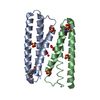
Mass: 96.063 Da / Num. of mol.: 5 / Source method: obtained synthetically / Formula: SO4
#4: Water ChemComp-HOH / water


Mass: 18.015 Da / Num. of mol.: 174 / Source method: isolated from a natural source / Formula: H2O
Has protein modificationY
Sequence detailsTHE CONSTRUCT (RESIDUES 23-143) WAS EXPRESSED WITH A PURIFICATION TAG MGSDKIHHHHHHENLYFQG. THE TAG ...THE CONSTRUCT (RESIDUES 23-143) WAS EXPRESSED WITH A PURIFICATION TAG MGSDKIHHHHHHENLYFQG. THE TAG WAS REMOVED WITH TEV PROTEASE LEAVING ONLY A GLYCINE (0) FOLLOWED BY THE TARGET SEQUENCE.

-
Experimental details

-
Experiment

ExperimentMethod: X-RAY DIFFRACTION / Number of used crystals: 1

-
Sample preparation

CrystalDensity Matthews: 2.64 Å3/Da / Density % sol: 53.47 %
Crystal growTemperature: 293 K / Method: vapor diffusion, sitting drop / pH: 5.67
Details: NANODROP, 2.10M Ammonium sulfate, 0.250M Potassium sodium tartrate, 0.1M Sodium citrate - citric acid pH 5.67, VAPOR DIFFUSION, SITTING DROP, temperature 293K

-
Data collection

DiffractionMean temperature: 100 K
Diffraction sourceSource: SYNCHROTRON / Site: SSRL / Beamline: BL9-2 / Wavelength: 0.91162, 0.97964, 0.97949
DetectorType: MARMOSAIC 325 mm CCD / Detector: CCD / Date: Jan 19, 2009 / Details: Flat collimating mirror, toroid focusing mirror
RadiationMonochromator: Double crystal / Protocol: MAD / Monochromatic (M) / Laue (L): M / Scattering type: x-ray
Radiation wavelength
IDWavelength (Å)Relative weight
10.911621
20.979641
30.979491
ReflectionResolution: 1.85→29.173 Å / Num. obs: 25875 / % possible obs: 100 % / Redundancy: 7.2 % / Biso Wilson estimate: 31.628 Å2 / Rmerge(I) obs: 0.065 / Rrim(I) all: 0.07 / Rsym value: 0.065 / Net I/av σ(I): 7.149 / Net I/σ(I): 18.5
Reflection shell

Diffraction-ID: 1

Resolution (Å)Redundancy (%)Rmerge(I) obsMean I/σ(I) obsNum. measured allNum. unique allRsym value% possible all
1.85-1.97.30.6922.71377418800.692100
1.9-1.957.30.5731.41334918180.573100
1.95-2.017.40.3712.11315717880.371100
2.01-2.077.30.2832.71270417390.283100
2.07-2.147.30.2273.41229916740.227100
2.14-2.217.30.1764.31181816150.176100
2.21-2.297.30.1345.61162215880.134100
2.29-2.397.30.1076.81108515130.107100
2.39-2.497.30.0997.21059714530.099100
2.49-2.627.30.0887.31024114040.088100
2.62-2.767.30.0847.7975113360.084100
2.76-2.937.30.0797.7921712690.079100
2.93-3.137.20.0678.6865012000.067100
3.13-3.387.30.05610.5802811070.056100
3.38-3.77.20.04612.6745310390.046100
3.7-4.147.20.04114.268449570.041100
4.14-4.7870.04812.558898370.048100
4.78-5.856.90.04812.149887270.048100
5.85-8.276.60.0371638795870.037100
8.27-29.1735.70.03417.619603440.03497.2

-
Phasing

PhasingMethod: MAD

-
Processing

Software
NameVersionClassificationNB
REFMAC5.5.0070refinement
PHENIXrefinement
SHELXphasing
MolProbity3beta29model building
SCALA3.2.5data scaling
PDB_EXTRACT3.006data extraction
MAR345CCDdata collection
MOSFLMdata reduction
SHELXDphasing
autoSHARPphasing
RefinementMethod to determine structure: MAD / Resolution: 1.85→29.173 Å / Cor.coef. Fo:Fc: 0.96 / Cor.coef. Fo:Fc free: 0.953 / Occupancy max: 1 / Occupancy min: 0.25 / SU B: 4.871 / SU ML: 0.066 / TLS residual ADP flag: LIKELY RESIDUAL / Cross valid method: THROUGHOUT / σ(F): 0 / ESU R: 0.114 / ESU R Free: 0.105
Stereochemistry target values: MAXIMUM LIKELIHOOD WITH PHASES
Details: 1. HYDROGENS HAVE BEEN ADDED IN THE RIDING POSITIONS. 2. A MET-INHIBITION PROTOCOL WAS USED FOR SELENOMETHIONINE INCORPORATION DURING PROTEIN EXPRESSION. THE OCCUPANCY OF THE SE ATOMS IN THE ...Details: 1. HYDROGENS HAVE BEEN ADDED IN THE RIDING POSITIONS. 2. A MET-INHIBITION PROTOCOL WAS USED FOR SELENOMETHIONINE INCORPORATION DURING PROTEIN EXPRESSION. THE OCCUPANCY OF THE SE ATOMS IN THE MSE RESIDUES WAS REDUCED TO 0.75 FOR THE REDUCED SCATTERING POWER DUE TO PARTIAL S-MET INCORPORATION. 3. ATOM RECORDS CONTAIN RESIDUAL B FACTORS ONLY. 4. SULFATE (SO4) MODELED IS PRESENT IN CRYSTALLIZATION CONDITIONS. 5. AN UNKNOWN LIGAND (UNL) WAS MODELED NEAR THE CONSERVED RESIDUE ARG 120. THE DENSITY IS DISORDERED IN THIS REGION.
RfactorNum. reflection% reflectionSelection details
Rfree0.199 1314 5.1 %RANDOM
Rwork0.178 24513 --
obs0.179 25827 99.96 %-
Solvent computationIon probe radii: 0.8 Å / Shrinkage radii: 0.8 Å / VDW probe radii: 1.2 Å / Solvent model: MASK
Displacement parametersBiso max: 88.78 Å2 / Biso mean: 25.285 Å2 / Biso min: 5.24 Å2
Baniso -1Baniso -2Baniso -3
1-0.17 Å20 Å20 Å2
2--0.17 Å20 Å2
3----0.33 Å2
Refinement stepCycle: LAST / Resolution: 1.85→29.173 Å
ProteinNucleic acidLigandSolventTotal
Num. atoms1561 0 33 174 1768
Refine LS restraints
Refine-IDTypeDev idealDev ideal targetNumber
X-RAY DIFFRACTIONr_bond_refined_d0.0180.0211791
X-RAY DIFFRACTIONr_bond_other_d0.0070.021240
X-RAY DIFFRACTIONr_angle_refined_deg1.4861.9442454
X-RAY DIFFRACTIONr_angle_other_deg1.1783.0033007
X-RAY DIFFRACTIONr_dihedral_angle_1_deg4.2355244
X-RAY DIFFRACTIONr_dihedral_angle_2_deg33.53624.364110
X-RAY DIFFRACTIONr_dihedral_angle_3_deg12.44115320
X-RAY DIFFRACTIONr_dihedral_angle_4_deg19.6681522
X-RAY DIFFRACTIONr_chiral_restr0.1510.2252
X-RAY DIFFRACTIONr_gen_planes_refined0.0060.022113
X-RAY DIFFRACTIONr_gen_planes_other0.0010.02389
X-RAY DIFFRACTIONr_mcbond_it1.79831078
X-RAY DIFFRACTIONr_mcbond_other0.7173433
X-RAY DIFFRACTIONr_mcangle_it2.86251730
X-RAY DIFFRACTIONr_scbond_it5.4238713
X-RAY DIFFRACTIONr_scangle_it7.98111699
Refine LS restraints NCS

Dom-ID: 1 / Auth asym-ID: A / Ens-ID: 1 / Refine-ID: X-RAY DIFFRACTION

NumberTypeRms dev position (Å)Weight position
580MEDIUM POSITIONAL0.350.5
755LOOSE POSITIONAL0.625
580MEDIUM THERMAL1.212
755LOOSE THERMAL1.3210
LS refinement shellResolution: 1.85→1.898 Å / Total num. of bins used: 20
RfactorNum. reflection% reflection
Rfree0.338 106 -
Rwork0.279 1771 -
all-1877 -
obs--100 %
Refinement TLS params.

Method: refined / Refine-ID: X-RAY DIFFRACTION

IDL112)L122)L132)L222)L232)L332)S11 (Å °)S12 (Å °)S13 (Å °)S21 (Å °)S22 (Å °)S23 (Å °)S31 (Å °)S32 (Å °)S33 (Å °)T112)T122)T132)T222)T232)T332)Origin x (Å)Origin y (Å)Origin z (Å)
11.43650.5577-0.91961.2344-0.33691.34590.013-0.10540.02620.1760.0322-0.24170.0182-0.0662-0.04520.04060.0176-0.03480.0586-0.0070.069435.565725.29986.9905
22.69251.1379-0.71632.0696-0.521.67070.00490.0622-0.1867-0.0890.1081-0.07610.0112-0.0875-0.1130.03440.00970.01480.01510.010.039126.273310.9451-0.8596
Refinement TLS group
IDRefine-IDRefine TLS-IDAuth asym-IDAuth seq-ID
1X-RAY DIFFRACTION1A33 - 135
2X-RAY DIFFRACTION2B37 - 134

+
About Yorodumi

-
News

-
Feb 9, 2022. New format data for meta-information of EMDB entries

New format data for meta-information of EMDB entries

  • Version 3 of the EMDB header file is now the official format.
  • The previous official version 1.9 will be removed from the archive.

Related info.:EMDB header

External links:wwPDB to switch to version 3 of the EMDB data model

-
Aug 12, 2020. Covid-19 info

Covid-19 info

URL: https://pdbj.org/emnavi/covid19.php

New page: Covid-19 featured information page in EM Navigator.

Related info.:Covid-19 info / Mar 5, 2020. Novel coronavirus structure data

+
Mar 5, 2020. Novel coronavirus structure data

Novel coronavirus structure data

Related info.:Yorodumi Speices / Aug 12, 2020. Covid-19 info

External links:COVID-19 featured content - PDBj / Molecule of the Month (242):Coronavirus Proteases

+
Jan 31, 2019. EMDB accession codes are about to change! (news from PDBe EMDB page)

EMDB accession codes are about to change! (news from PDBe EMDB page)

  • The allocation of 4 digits for EMDB accession codes will soon come to an end. Whilst these codes will remain in use, new EMDB accession codes will include an additional digit and will expand incrementally as the available range of codes is exhausted. The current 4-digit format prefixed with “EMD-” (i.e. EMD-XXXX) will advance to a 5-digit format (i.e. EMD-XXXXX), and so on. It is currently estimated that the 4-digit codes will be depleted around Spring 2019, at which point the 5-digit format will come into force.
  • The EM Navigator/Yorodumi systems omit the EMD- prefix.

Related info.:Q: What is EMD? / ID/Accession-code notation in Yorodumi/EM Navigator

External links:EMDB Accession Codes are Changing Soon! / Contact to PDBj

+
Jul 12, 2017. Major update of PDB

Major update of PDB

  • wwPDB released updated PDB data conforming to the new PDBx/mmCIF dictionary.
  • This is a major update changing the version number from 4 to 5, and with Remediation, in which all the entries are updated.
  • In this update, many items about electron microscopy experimental information are reorganized (e.g. em_software).
  • Now, EM Navigator and Yorodumi are based on the updated data.

External links:wwPDB Remediation / Enriched Model Files Conforming to OneDep Data Standards Now Available in the PDB FTP Archive

-
Yorodumi

Thousand views of thousand structures

  • Yorodumi is a browser for structure data from EMDB, PDB, SASBDB, etc.
  • This page is also the successor to EM Navigator detail page, and also detail information page/front-end page for Omokage search.
  • The word "yorodu" (or yorozu) is an old Japanese word meaning "ten thousand". "mi" (miru) is to see.

Related info.:EMDB / PDB / SASBDB / Comparison of 3 databanks / Yorodumi Search / Aug 31, 2016. New EM Navigator & Yorodumi / Yorodumi Papers / Jmol/JSmol / Function and homology information / Changes in new EM Navigator and Yorodumi

Read more