 Movie
Movie Controller

[English] 日本語
 Yorodumi
- PDB-1nog: Crystal Structure of Conserved Protein 0546 from Thermoplasma Aci... -
+ Open data

- Basic information
| Entry | Database: PDB / ID: 1nog | ||||||
|---|---|---|---|---|---|---|---|
| Title | Crystal Structure of Conserved Protein 0546 from Thermoplasma Acidophilum | ||||||
 Components Components | conserved hypothetical protein TA0546 | ||||||
 Keywords Keywords | STRUCTURAL GENOMICS / UNKNOWN FUNCTION / PSI / Protein Structure Initiative / Midwest Center for Structural Genomics / MCSG | ||||||
| Function / homology |  Function and homology information Function and homology information | ||||||
| Biological species |   Thermoplasma acidophilum (acidophilic) Thermoplasma acidophilum (acidophilic) | ||||||
| Method |  X-RAY DIFFRACTION / SYNCHROTRON / MAD / Resolution: 1.55 Å X-RAY DIFFRACTION / SYNCHROTRON / MAD / Resolution: 1.55 Å | ||||||
 Authors Authors | Saridakis, V. / Sanishvili, R. / Iakounine, A. / Xu, X. / Pennycooke, M. / Gu, J. / Joachimiak, A. / Arrowsmith, C.H. / Edwards, A.M. / Christendat, D. / Midwest Center for Structural Genomics (MCSG) | ||||||
 Citation Citation | Journal: J.Biol.Chem. / Year: 2004 Title: The structural basis for methylmalonic aciduria. The crystal structure of archaeal ATP:cobalamin adenosyltransferase. Authors: Saridakis, V. / Yakunin, A. / Xu, X. / Anandakumar, P. / Pennycooke, M. / Gu, J. / Cheung, F. / Lew, J.M. / Sanishvili, R. / Joachimiak, A. / Arrowsmith, C.H. / Christendat, D. / Edwards, A.M. | ||||||
| History |
| ||||||
| Remark 999 | SEQUENCE The authors state that According to the density, the residue at position 110 is valine and ...SEQUENCE The authors state that According to the density, the residue at position 110 is valine and the residue at position 159 is ile. |
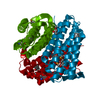
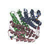
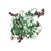
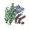
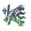
- Structure visualization
| Structure viewer | Molecule: MolmilJmol/JSmol |
|---|
- Downloads & links
-Download
| PDBx/mmCIF format | 1nog.cif.gz | 47.5 KB | Display | PDBx/mmCIF format |
|---|---|---|---|---|
| PDB format | pdb1nog.ent.gz | 34.4 KB | Display | PDB format |
| PDBx/mmJSON format | 1nog.json.gz | Tree view | PDBx/mmJSON format | |
| Others |  Other downloads Other downloads |
-Validation report
| Arichive directory | https://data.pdbj.org/pub/pdb/validation_reports/no/1nogftp://data.pdbj.org/pub/pdb/validation_reports/no/1nog | HTTPS FTP |
|---|
-Related structure data
| Similar structure data | |
|---|---|
| Other databases |


-Links
 PDBj
PDBj- Assembly
| Deposited unit | 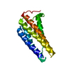
| ||||||||
|---|---|---|---|---|---|---|---|---|---|
| 1 | 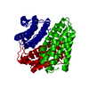
| ||||||||
| 2 | x 12
| ||||||||
| Unit cell |
| ||||||||
| Details | The biological assembly is a trimer generated from the monomer in the asymmetric unit by crystallographic symmetry. |
-Components
| #1: Protein | Mass: 20029.074 Da / Num. of mol.: 1 Source method: isolated from a genetically manipulated source Source: (gene. exp.) Thermoplasma acidophilum (acidophilic) / Gene: TA1434 / Plasmid: pET15b / Species (production host): Escherichia coli / Production host:  |
|---|---|
| #2: Water | ChemComp-HOH /  Mass: 18.015 Da / Num. of mol.: 170 / Source method: isolated from a natural source / Formula: H2O Mass: 18.015 Da / Num. of mol.: 170 / Source method: isolated from a natural source / Formula: H2O |
-Experimental details
-Experiment
| Experiment | Method: X-RAY DIFFRACTION / Number of used crystals: 1 |
|---|
- Sample preparation
| Crystal | Density Matthews: 2.94 Å3/Da / Density % sol: 58.18 % | |||||||||||||||
|---|---|---|---|---|---|---|---|---|---|---|---|---|---|---|---|---|
| Crystal grow | Temperature: 295 K / Method: vapor diffusion, hanging drop / pH: 7.5 Details: 0.4 M ammonium phosphate, 6 % methylpentanediol, pH 7.5, VAPOR DIFFUSION, HANGING DROP, temperature 295K | |||||||||||||||
| Crystal grow | *PLUS Temperature: 22 ℃ / pH: 4.6 / Method: vapor diffusion, hanging drop / Details: Christendat, D., (2000) J.Biol.Chem., 275, 24608. | |||||||||||||||
| Components of the solutions | *PLUS
|
-Data collection
| Diffraction | Mean temperature: 100 K |
|---|---|
| Diffraction source | Source: SYNCHROTRON / Site: APS  / Beamline: 19-ID / Beamline: 19-ID |
| Detector | Type: CUSTOM-MADE / Detector: CCD / Date: Feb 14, 2001 |
| Radiation | Protocol: MAD / Monochromatic (M) / Laue (L): M / Scattering type: x-ray |
| Radiation wavelength | Relative weight: 1 |
| Reflection | Resolution: 1.55→30 Å / Num. obs: 34479 / % possible obs: 99.9 % / Redundancy: 10.3 % / Biso Wilson estimate: 20 Å2 / Rmerge(I) obs: 0.101 / Net I/σ(I): 21.1 |
| Reflection shell | Resolution: 1.55→1.6 Å / Rmerge(I) obs: 0.504 / Mean I/σ(I) obs: 4.4 / Num. unique all: 3233 / % possible all: 100 |
| Reflection | *PLUS Num. obs: 62540 / % possible obs: 99 % / Num. measured all: 341835 / Rmerge(I) obs: 0.094 |
| Reflection shell | *PLUS % possible obs: 99.8 % / Rmerge(I) obs: 0.47 |
- Processing
| Software |
| ||||||||||||||||||||||||||||||||||||
|---|---|---|---|---|---|---|---|---|---|---|---|---|---|---|---|---|---|---|---|---|---|---|---|---|---|---|---|---|---|---|---|---|---|---|---|---|---|
| Refinement | Method to determine structure: MAD / Resolution: 1.55→29.69 Å / Rfactor Rfree error: 0.006 / Isotropic thermal model: RESTRAINED / Cross valid method: THROUGHOUT / σ(F): 0
| ||||||||||||||||||||||||||||||||||||
| Solvent computation | Solvent model: FLAT MODEL / Bsol: 51.7177 Å2 / ksol: 0.378784 e/Å3 | ||||||||||||||||||||||||||||||||||||
| Displacement parameters | Biso mean: 23.1 Å2
| ||||||||||||||||||||||||||||||||||||
| Refine analyze | Luzzati coordinate error free: 0.19 Å / Luzzati sigma a free: 0.13 Å | ||||||||||||||||||||||||||||||||||||
| Refinement step | Cycle: LAST / Resolution: 1.55→29.69 Å
| ||||||||||||||||||||||||||||||||||||
| Refine LS restraints |
| ||||||||||||||||||||||||||||||||||||
| LS refinement shell | Resolution: 1.55→1.65 Å / Rfactor Rfree error: 0.015 / Total num. of bins used: 6
| ||||||||||||||||||||||||||||||||||||
| Xplor file |
| ||||||||||||||||||||||||||||||||||||
| Refine LS restraints | *PLUS
|
