- PDB-5vxm: 2.05 A resolution structure of IpaD from Shigella flexneri in com... -
+
Open data
ID or keywords:
Loading...
-
Basic information
Entry
Database: PDB / ID: 5vxm
Title
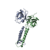
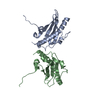
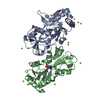
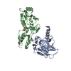
2.05 A resolution structure of IpaD from Shigella flexneri in complex with single-domain antibody 20ipaD
Components
Invasin IpaD
Single-domain antibody 20ipaD
Keywords
IMMUNE SYSTEM / tip protein / VHH / T3SS
Function / homology
Function and homology information
effector-mediated activation of programmed cell death in host / extracellular region Similarity search - Function
IpaD-like / IpaD-like / Type III secretion systems tip complex components / BipD-like superfamily / Type III secretion systems tip complex components / Up-down Bundle / Mainly Alpha Similarity search - Domain/homology
Mass: 31904.623 Da / Num. of mol.: 1 / Mutation: C322S Source method: isolated from a genetically manipulated source Source: (gene. exp.) Shigella flexneri (bacteria) / Gene: ipaD, CP0126 / Production host: Escherichia coli (E. coli) / References: UniProt: P18013
#2: Antibody
Single-domainantibody20ipaD
Mass: 16253.950 Da / Num. of mol.: 1 Source method: isolated from a genetically manipulated source Source: (gene. exp.) Vicugna pacos (alpaca) / Production host: Escherichia coli (E. coli)
In the structure databanks used in Yorodumi, some data are registered as the other names, "COVID-19 virus" and "2019-nCoV". Here are the details of the virus and the list of structure data.
Jan 31, 2019. EMDB accession codes are about to change! (news from PDBe EMDB page)
EMDB accession codes are about to change! (news from PDBe EMDB page)
The allocation of 4 digits for EMDB accession codes will soon come to an end. Whilst these codes will remain in use, new EMDB accession codes will include an additional digit and will expand incrementally as the available range of codes is exhausted. The current 4-digit format prefixed with “EMD-” (i.e. EMD-XXXX) will advance to a 5-digit format (i.e. EMD-XXXXX), and so on. It is currently estimated that the 4-digit codes will be depleted around Spring 2019, at which point the 5-digit format will come into force.
The EM Navigator/Yorodumi systems omit the EMD- prefix.
Related info.:Q: What is EMD? / ID/Accession-code notation in Yorodumi/EM Navigator
Yorodumi is a browser for structure data from EMDB, PDB, SASBDB, etc.
This page is also the successor to EM Navigator detail page, and also detail information page/front-end page for Omokage search.
The word "yorodu" (or yorozu) is an old Japanese word meaning "ten thousand". "mi" (miru) is to see.
Related info.:EMDB / PDB / SASBDB / Comparison of 3 databanks / Yorodumi Search / Aug 31, 2016. New EM Navigator & Yorodumi / Yorodumi Papers / Jmol/JSmol / Function and homology information / Changes in new EM Navigator and Yorodumi
 Movie
Movie Controller

 Yorodumi
 Open data
 Basic information
 Components
 Keywords
 Function and homology information
 Shigella flexneri (bacteria)
 X-RAY DIFFRACTION /
  Authors
 Citation
 Structure visualization
 Downloads & links
 Other downloads
 PDBj
PDBj

 Assembly
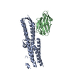
 Mass: 18.015 Da / Num. of mol.: 68 / Source method: isolated from a natural source / Formula: H2O
 Sample preparation
 / Beamline: 17-ID / Wavelength: 1 Å
 Processing