 Movie
Movie Controller

[English] 日本語
 Yorodumi
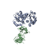
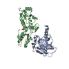
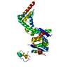
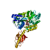
- PDB-3wvz: Crystal structure of Hikeshi, a new nuclear transport receptor of... -
+ Open data

- Basic information
| Entry | Database: PDB / ID: 3wvz | ||||||
|---|---|---|---|---|---|---|---|
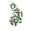
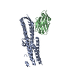
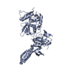
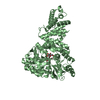
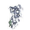
| Title | Crystal structure of Hikeshi, a new nuclear transport receptor of Hsp70 | ||||||
 Components Components | Protein Hikeshi | ||||||
 Keywords Keywords | TRANSPORT PROTEIN / Nuclear transport receptor | ||||||
| Function / homology |  Function and homology information Function and homology informationnuclear import signal receptor activity / Regulation of HSF1-mediated heat shock response / Hsp70 protein binding / protein import into nucleus / protein transport / cellular response to heat / nucleoplasm / nucleus / cytosol Similarity search - Function | ||||||
| Biological species |  Homo sapiens (human) Homo sapiens (human) | ||||||
| Method |  X-RAY DIFFRACTION / SYNCHROTRON / MOLECULAR REPLACEMENT / Resolution: 1.88 Å X-RAY DIFFRACTION / SYNCHROTRON / MOLECULAR REPLACEMENT / Resolution: 1.88 Å | ||||||
 Authors Authors | Song, J. / Kose, S. / Watanabe, A. / Son, S.Y. / Choi, S. / Hong, R.H. / Yamashita, E. / Park, I.Y. / Imamoto, N. / Lee, S.J. | ||||||
 Citation Citation | Journal: Acta Crystallogr.,Sect.D / Year: 2015 Title: Structural and functional analysis of Hikeshi, a new nuclear transport receptor of Hsp70s Authors: Song, J. / Kose, S. / Watanabe, A. / Son, S.Y. / Choi, S. / Hong, H. / Yamashita, E. / Park, I.Y. / Imamoto, N. / Lee, S.J. | ||||||
| History |
|
- Structure visualization
| Structure viewer | Molecule: MolmilJmol/JSmol |
|---|
- Downloads & links
-Download
| PDBx/mmCIF format | 3wvz.cif.gz | 160.5 KB | Display | PDBx/mmCIF format |
|---|---|---|---|---|
| PDB format | pdb3wvz.ent.gz | 128.4 KB | Display | PDB format |
| PDBx/mmJSON format | 3wvz.json.gz | Tree view | PDBx/mmJSON format | |
| Others |  Other downloads Other downloads |
-Validation report
| Arichive directory | https://data.pdbj.org/pub/pdb/validation_reports/wv/3wvzftp://data.pdbj.org/pub/pdb/validation_reports/wv/3wvz | HTTPS FTP |
|---|
-Related structure data


-Links
 PDBj
PDBj

- Assembly
| Deposited unit | 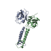
| |||||||||
|---|---|---|---|---|---|---|---|---|---|---|
| 1 |
| |||||||||
| Unit cell |
| |||||||||
| Components on special symmetry positions |
|
-Components
| #1: Protein | Mass: 21968.982 Da / Num. of mol.: 2 Source method: isolated from a genetically manipulated source Source: (gene. exp.) Homo sapiens (human) / Gene: C11orf73, HSPC138, HSPC179, HSPC248 / Production host:  #2: Water | ChemComp-HOH / |  Mass: 18.015 Da / Num. of mol.: 226 / Source method: isolated from a natural source / Formula: H2O Mass: 18.015 Da / Num. of mol.: 226 / Source method: isolated from a natural source / Formula: H2O |
|---|
-Experimental details
-Experiment
| Experiment | Method: X-RAY DIFFRACTION / Number of used crystals: 1 |
|---|
- Sample preparation
| Crystal | Density Matthews: 2.35 Å3/Da / Density % sol: 47.6 % |
|---|---|
| Crystal grow | Temperature: 293.15 K / Method: vapor diffusion, hanging drop / pH: 7.4 Details: 0.1M Tris-HCl (pH 7.4), 1M ammonium sulfate, VAPOR DIFFUSION, HANGING DROP, temperature 293.15K |
-Data collection
| Diffraction | Mean temperature: 90 K |
|---|---|
| Diffraction source | Source: SYNCHROTRON / Site: SPring-8  / Beamline: BL44XU / Wavelength: 0.9192 Å / Beamline: BL44XU / Wavelength: 0.9192 Å |
| Detector | Detector: AREA DETECTOR / Date: Nov 8, 2013 / Details: 0.9192 eV |
| Radiation | Protocol: SINGLE WAVELENGTH / Monochromatic (M) / Laue (L): M / Scattering type: x-ray |
| Radiation wavelength | Wavelength: 0.9192 Å / Relative weight: 1 |
| Reflection | Resolution: 1.88→100 Å / Num. all: 32074 / Num. obs: 19445 |
| Reflection shell | Resolution: 1.88→100 Å |
- Processing
| Software |
| ||||||||||||||||||||||||||||||||||||||||||||||||||||||||||||||||||||||||||||||
|---|---|---|---|---|---|---|---|---|---|---|---|---|---|---|---|---|---|---|---|---|---|---|---|---|---|---|---|---|---|---|---|---|---|---|---|---|---|---|---|---|---|---|---|---|---|---|---|---|---|---|---|---|---|---|---|---|---|---|---|---|---|---|---|---|---|---|---|---|---|---|---|---|---|---|---|---|---|---|---|
| Refinement | Method to determine structure: MOLECULAR REPLACEMENT / Resolution: 1.88→39.909 Å / SU ML: 0.22 / σ(F): 1.35 / Phase error: 23.6 / Stereochemistry target values: ML
| ||||||||||||||||||||||||||||||||||||||||||||||||||||||||||||||||||||||||||||||
| Solvent computation | Shrinkage radii: 0.9 Å / VDW probe radii: 1.11 Å / Solvent model: FLAT BULK SOLVENT MODEL | ||||||||||||||||||||||||||||||||||||||||||||||||||||||||||||||||||||||||||||||
| Refinement step | Cycle: LAST / Resolution: 1.88→39.909 Å
| ||||||||||||||||||||||||||||||||||||||||||||||||||||||||||||||||||||||||||||||
| Refine LS restraints |
| ||||||||||||||||||||||||||||||||||||||||||||||||||||||||||||||||||||||||||||||
| LS refinement shell | Refine-ID: X-RAY DIFFRACTION / Total num. of bins used: 12
| ||||||||||||||||||||||||||||||||||||||||||||||||||||||||||||||||||||||||||||||
| Refinement TLS params. | Method: refined / Origin x: 13.6688 Å / Origin y: 25.8113 Å / Origin z: 95.1139 Å
| ||||||||||||||||||||||||||||||||||||||||||||||||||||||||||||||||||||||||||||||
| Refinement TLS group | Selection details: all |
