 Movie
Movie Controller

[English] 日本語
 Yorodumi
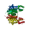
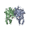
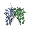
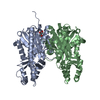
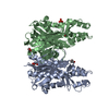
- PDB-5vxe: Crystal structure of Xanthomonas campestris OleA E117A bound with... -
+ Open data

- Basic information
| Entry | Database: PDB / ID: 5vxe | ||||||
|---|---|---|---|---|---|---|---|
| Title | Crystal structure of Xanthomonas campestris OleA E117A bound with Cerulenin | ||||||
 Components Components | 3-oxoacyl-[ACP] synthase III | ||||||
 Keywords Keywords | TRANSFERASE / Thiolase / Condensation | ||||||
| Function / homology |  Function and homology information Function and homology informationacyl-CoA:acyl-CoA alkyltransferase / secondary metabolite biosynthetic process / 3-oxoacyl-[acyl-carrier-protein] synthase activity / fatty acid biosynthetic process / metal ion binding / cytoplasm Similarity search - Function | ||||||
| Biological species |  Xanthomonas campestris pv. campestris (bacteria) Xanthomonas campestris pv. campestris (bacteria) | ||||||
| Method |  X-RAY DIFFRACTION / SYNCHROTRON / MOLECULAR REPLACEMENT / Resolution: 1.66 Å X-RAY DIFFRACTION / SYNCHROTRON / MOLECULAR REPLACEMENT / Resolution: 1.66 Å | ||||||
 Authors Authors | Jensen, M.R. / Wilmot, C.M. | ||||||
| Funding support |  United States, 1items United States, 1items
| ||||||
 Citation Citation | Journal: Biochem. J. / Year: 2017 Title: OleA Glu117 is key to condensation of two fatty-acyl coenzyme A substrates in long-chain olefin biosynthesis. Authors: Jensen, M.R. / Goblirsch, B.R. / Christenson, J.K. / Esler, M.A. / Mohamed, F.A. / Wackett, L.P. / Wilmot, C.M. | ||||||
| History |
|
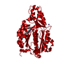
- Structure visualization
| Structure viewer | Molecule: MolmilJmol/JSmol |
|---|
- Downloads & links
-Download
| PDBx/mmCIF format | 5vxe.cif.gz | 149.1 KB | Display | PDBx/mmCIF format |
|---|---|---|---|---|
| PDB format | pdb5vxe.ent.gz | 115 KB | Display | PDB format |
| PDBx/mmJSON format | 5vxe.json.gz | Tree view | PDBx/mmJSON format | |
| Others |  Other downloads Other downloads |
-Validation report
| Arichive directory | https://data.pdbj.org/pub/pdb/validation_reports/vx/5vxeftp://data.pdbj.org/pub/pdb/validation_reports/vx/5vxe | HTTPS FTP |
|---|
-Related structure data
| Related structure data |  5vxdC 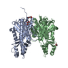 5vxfC  5vxgC 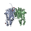 5vxhC 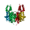 5vxiC 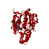 3s21S C: citing same article ( S: Starting model for refinement |
|---|---|
| Similar structure data |


-Links
 PDBj
PDBj

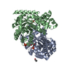
- Assembly
| Deposited unit | 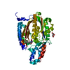
| ||||||||
|---|---|---|---|---|---|---|---|---|---|
| 1 | 
| ||||||||
| Unit cell |
|
-Components
| #1: Protein | Mass: 38784.406 Da / Num. of mol.: 1 / Mutation: E117A Source method: isolated from a genetically manipulated source Source: (gene. exp.) Xanthomonas campestris pv. campestris (strain ATCC 33913 / DSM 3586 / NCPPB 528 / LMG 568 / P 25) (bacteria)Strain: ATCC 33913 / DSM 3586 / NCPPB 528 / LMG 568 / P 25 / Gene: fabH, XCC0212 / Plasmid: pET28b+ / Production host: |
|---|---|
| #2: Chemical | ChemComp-CER / (  Mass: 225.284 Da / Num. of mol.: 1 / Source method: obtained synthetically / Formula: C12H19NO3 / Comment: antifungal, antibiotic*YM Mass: 225.284 Da / Num. of mol.: 1 / Source method: obtained synthetically / Formula: C12H19NO3 / Comment: antifungal, antibiotic*YM |
| #3: Chemical | ChemComp-GOL /   Mass: 92.094 Da / Num. of mol.: 1 / Source method: obtained synthetically / Formula: C3H8O3 Mass: 92.094 Da / Num. of mol.: 1 / Source method: obtained synthetically / Formula: C3H8O3 |
| #4: Chemical | ChemComp-MN /   Mass: 54.938 Da / Num. of mol.: 1 / Source method: obtained synthetically / Formula: Mn Mass: 54.938 Da / Num. of mol.: 1 / Source method: obtained synthetically / Formula: Mn |
| #5: Water | ChemComp-HOH /  Mass: 18.015 Da / Num. of mol.: 109 / Source method: isolated from a natural source / Formula: H2O Mass: 18.015 Da / Num. of mol.: 109 / Source method: isolated from a natural source / Formula: H2O |
-Experimental details
-Experiment
| Experiment | Method: X-RAY DIFFRACTION / Number of used crystals: 1 |
|---|
- Sample preparation
| Crystal | Density Matthews: 2.26 Å3/Da / Density % sol: 41.31 % / Mosaicity: 0.313 ° |
|---|---|
| Crystal grow | Temperature: 292 K / Method: vapor diffusion, hanging drop / pH: 6 Details: 13% PEG 4000, 100 MM MANGANESE CHLORIDE, 100 MM MES PH 6.0 |
-Data collection
| Diffraction | Mean temperature: 100 K | |||||||||||||||||||||||||||||||||||||||||||||||||||||||||||||||||||||||||||||||||||||||||||||||||||||||||||||||||||||||||||||||||||||||||||||||||||||||||||||||||||||||||||||||||||||||||||||
|---|---|---|---|---|---|---|---|---|---|---|---|---|---|---|---|---|---|---|---|---|---|---|---|---|---|---|---|---|---|---|---|---|---|---|---|---|---|---|---|---|---|---|---|---|---|---|---|---|---|---|---|---|---|---|---|---|---|---|---|---|---|---|---|---|---|---|---|---|---|---|---|---|---|---|---|---|---|---|---|---|---|---|---|---|---|---|---|---|---|---|---|---|---|---|---|---|---|---|---|---|---|---|---|---|---|---|---|---|---|---|---|---|---|---|---|---|---|---|---|---|---|---|---|---|---|---|---|---|---|---|---|---|---|---|---|---|---|---|---|---|---|---|---|---|---|---|---|---|---|---|---|---|---|---|---|---|---|---|---|---|---|---|---|---|---|---|---|---|---|---|---|---|---|---|---|---|---|---|---|---|---|---|---|---|---|---|---|---|---|---|
| Diffraction source | Source: SYNCHROTRON / Site: APS / Beamline: 23-ID-B / Wavelength: 1.03317 Å | |||||||||||||||||||||||||||||||||||||||||||||||||||||||||||||||||||||||||||||||||||||||||||||||||||||||||||||||||||||||||||||||||||||||||||||||||||||||||||||||||||||||||||||||||||||||||||||
| Detector | Type: MAR scanner 300 mm plate / Detector: IMAGE PLATE / Date: Jun 14, 2015 | |||||||||||||||||||||||||||||||||||||||||||||||||||||||||||||||||||||||||||||||||||||||||||||||||||||||||||||||||||||||||||||||||||||||||||||||||||||||||||||||||||||||||||||||||||||||||||||
| Radiation | Monochromator: SI 111 CHANNEL / Protocol: SINGLE WAVELENGTH / Monochromatic (M) / Laue (L): M / Scattering type: x-ray | |||||||||||||||||||||||||||||||||||||||||||||||||||||||||||||||||||||||||||||||||||||||||||||||||||||||||||||||||||||||||||||||||||||||||||||||||||||||||||||||||||||||||||||||||||||||||||||
| Radiation wavelength | Wavelength: 1.03317 Å / Relative weight: 1 | |||||||||||||||||||||||||||||||||||||||||||||||||||||||||||||||||||||||||||||||||||||||||||||||||||||||||||||||||||||||||||||||||||||||||||||||||||||||||||||||||||||||||||||||||||||||||||||
| Reflection | Resolution: 1.66→50 Å / Num. obs: 38595 / % possible obs: 99.3 % / Redundancy: 5.6 % / Biso Wilson estimate: 21.7 Å2 / Rmerge(I) obs: 0.077 / Rpim(I) all: 0.035 / Rrim(I) all: 0.085 / Χ2: 1.053 / Net I/av σ(I): 21.6 / Net I/σ(I): 10.7 | |||||||||||||||||||||||||||||||||||||||||||||||||||||||||||||||||||||||||||||||||||||||||||||||||||||||||||||||||||||||||||||||||||||||||||||||||||||||||||||||||||||||||||||||||||||||||||||
| Reflection shell | Diffraction-ID: 1
|
- Processing
| Software |
| ||||||||||||||||||||||||||||||||||||||||||||||||||||||||||||
|---|---|---|---|---|---|---|---|---|---|---|---|---|---|---|---|---|---|---|---|---|---|---|---|---|---|---|---|---|---|---|---|---|---|---|---|---|---|---|---|---|---|---|---|---|---|---|---|---|---|---|---|---|---|---|---|---|---|---|---|---|---|
| Refinement | Method to determine structure: MOLECULAR REPLACEMENT Starting model: 3S21 Resolution: 1.66→50 Å / Cor.coef. Fo:Fc: 0.968 / Cor.coef. Fo:Fc free: 0.953 / SU B: 4.45 / SU ML: 0.073 / Cross valid method: THROUGHOUT / σ(F): 0 / ESU R: 0.095 / ESU R Free: 0.096 / Details: HYDROGENS HAVE BEEN ADDED IN THE RIDING POSITIONS
| ||||||||||||||||||||||||||||||||||||||||||||||||||||||||||||
| Solvent computation | Ion probe radii: 0.8 Å / Shrinkage radii: 0.8 Å / VDW probe radii: 1.2 Å | ||||||||||||||||||||||||||||||||||||||||||||||||||||||||||||
| Displacement parameters | Biso max: 107.14 Å2 / Biso mean: 34.301 Å2 / Biso min: 11.77 Å2
| ||||||||||||||||||||||||||||||||||||||||||||||||||||||||||||
| Refinement step | Cycle: final / Resolution: 1.66→50 Å
| ||||||||||||||||||||||||||||||||||||||||||||||||||||||||||||
| Refine LS restraints |
| ||||||||||||||||||||||||||||||||||||||||||||||||||||||||||||
| LS refinement shell | Resolution: 1.658→1.701 Å / Rfactor Rfree error: 0 / Total num. of bins used: 20
| ||||||||||||||||||||||||||||||||||||||||||||||||||||||||||||
| Refinement TLS params. | Method: refined / Origin x: 20.6023 Å / Origin y: -36.873 Å / Origin z: 8.0781 Å
|
