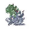
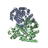
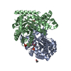
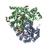
Journal: To be Published Title: Crystal structure of Plasmodium falciparum orotidine 5'-monophosphate decarboxylase covalently modified by 2-prime-fluoro-6-iodo-UMP Authors: Liu, Y. / Kotra, L.P. / Pai, E.F.
Type: MARMOSAIC 225 mm CCD / Detector: CCD / Date: Aug 16, 2007 Details: DCM with cryo-cooled 1st crystal sagittally bent 2nd crystal followed by vertically focusing mirror.
Radiation
Protocol: SINGLE WAVELENGTH / Monochromatic (M) / Laue (L): M / Scattering type: x-ray
Resolution: 1.75→50 Å / Cor.coef. Fo:Fc: 0.965 / Cor.coef. Fo:Fc free: 0.953 / SU B: 2.345 / SU ML: 0.076 / Cross valid method: THROUGHOUT / ESU R Free: 0.116 / Stereochemistry target values: MAXIMUM LIKELIHOOD / Details: HYDROGENS HAVE BEEN ADDED IN THE RIDING POSITIONS
Rfactor
Num. reflection
% reflection
Selection details
Rfree
0.19829
3035
5.1 %
RANDOM
Rwork
0.16229
-
-
-
obs
0.16413
56929
97.57 %
-
Solvent computation
Ion probe radii: 0.8 Å / Shrinkage radii: 0.8 Å / VDW probe radii: 1.4 Å / Solvent model: MASK
Displacement parameters
Biso mean: 17.807 Å2
Baniso -1
Baniso -2
Baniso -3
1-
0.29 Å2
0 Å2
0 Å2
2-
-
-0.02 Å2
0 Å2
3-
-
-
-0.27 Å2
Refinement step
Cycle: LAST / Resolution: 1.75→50 Å
Protein
Nucleic acid
Ligand
Solvent
Total
Num. atoms
5360
0
54
454
5868
Refine LS restraints
Refine-ID
Type
Dev ideal
Dev ideal target
Number
X-RAY DIFFRACTION
r_bond_refined_d
0.01
0.022
5735
X-RAY DIFFRACTION
r_angle_refined_deg
1.508
1.964
7764
X-RAY DIFFRACTION
r_dihedral_angle_1_deg
6.013
5
694
X-RAY DIFFRACTION
r_dihedral_angle_2_deg
38.423
25.864
295
X-RAY DIFFRACTION
r_dihedral_angle_3_deg
14.048
15
1056
X-RAY DIFFRACTION
r_dihedral_angle_4_deg
21.104
15
13
X-RAY DIFFRACTION
r_chiral_restr
0.109
0.2
822
X-RAY DIFFRACTION
r_gen_planes_refined
0.007
0.02
4397
X-RAY DIFFRACTION
r_nbd_refined
0.21
0.2
2984
X-RAY DIFFRACTION
r_nbtor_refined
0.316
0.2
4088
X-RAY DIFFRACTION
r_xyhbond_nbd_refined
0.152
0.2
454
X-RAY DIFFRACTION
r_symmetry_vdw_refined
0.202
0.2
101
X-RAY DIFFRACTION
r_symmetry_hbond_refined
0.216
0.2
24
X-RAY DIFFRACTION
r_mcbond_it
1.006
1.5
3471
X-RAY DIFFRACTION
r_mcangle_it
1.543
2
5518
X-RAY DIFFRACTION
r_scbond_it
2.678
3
2569
X-RAY DIFFRACTION
r_scangle_it
3.872
4.5
2246
LS refinement shell
Resolution: 1.751→1.796 Å / Total num. of bins used: 20
Rfactor
Num. reflection
% reflection
Rfree
0.301
234
-
Rwork
0.237
4036
-
obs
-
4270
95.93 %
+
About Yorodumi
-
News
-
Feb 9, 2022. New format data for meta-information of EMDB entries
New format data for meta-information of EMDB entries
Version 3 of the EMDB header file is now the official format.
The previous official version 1.9 will be removed from the archive.
In the structure databanks used in Yorodumi, some data are registered as the other names, "COVID-19 virus" and "2019-nCoV". Here are the details of the virus and the list of structure data.
Jan 31, 2019. EMDB accession codes are about to change! (news from PDBe EMDB page)
EMDB accession codes are about to change! (news from PDBe EMDB page)
The allocation of 4 digits for EMDB accession codes will soon come to an end. Whilst these codes will remain in use, new EMDB accession codes will include an additional digit and will expand incrementally as the available range of codes is exhausted. The current 4-digit format prefixed with “EMD-” (i.e. EMD-XXXX) will advance to a 5-digit format (i.e. EMD-XXXXX), and so on. It is currently estimated that the 4-digit codes will be depleted around Spring 2019, at which point the 5-digit format will come into force.
The EM Navigator/Yorodumi systems omit the EMD- prefix.
Related info.:Q: What is EMD? / ID/Accession-code notation in Yorodumi/EM Navigator
Yorodumi is a browser for structure data from EMDB, PDB, SASBDB, etc.
This page is also the successor to EM Navigator detail page, and also detail information page/front-end page for Omokage search.
The word "yorodu" (or yorozu) is an old Japanese word meaning "ten thousand". "mi" (miru) is to see.
Related info.:EMDB / PDB / SASBDB / Comparison of 3 databanks / Yorodumi Search / Aug 31, 2016. New EM Navigator & Yorodumi / Yorodumi Papers / Jmol/JSmol / Function and homology information / Changes in new EM Navigator and Yorodumi
 Movie
Movie Controller

 Yorodumi
 Open data
 Basic information
 Components
 Keywords
 Function and homology information
 X-RAY DIFFRACTION /
  Authors
 Citation
 Structure visualization
 Downloads & links
 Other downloads


 PDBj
PDBj
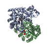
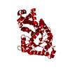
 Assembly
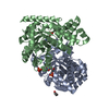

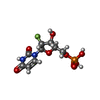
 Type: DNA linking / Mass: 326.172 Da / Num. of mol.: 2 / Source method: obtained synthetically / Formula: C9H12FN2O8P
 Mass: 106.120 Da / Num. of mol.: 1 / Source method: obtained synthetically / Formula: C4H10O3
 Mass: 94.971 Da / Num. of mol.: 1 / Source method: obtained synthetically / Formula: PO4
 Mass: 18.015 Da / Num. of mol.: 454 / Source method: isolated from a natural source / Formula: H2O
 Sample preparation
 / Beamline: 08ID-1 / Wavelength: 0.97934 Å
 Processing