 Movie
Movie Controller

[English] 日本語
 Yorodumi
- PDB-3s9y: Crystal Structure of P. falciparum orotidine 5'-monophosphate dec... -
+ Open data

- Basic information
| Entry | Database: PDB / ID: 3s9y | ||||||
|---|---|---|---|---|---|---|---|
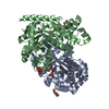
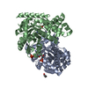
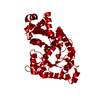
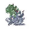
| Title | Crystal Structure of P. falciparum orotidine 5'-monophosphate decarboxylase complexed with 5-fluoro-6-amino-UMP in space group P21, produced from 5-fluoro-6-azido-UMP | ||||||
 Components Components | Orotidine 5'-phosphate decarboxylase | ||||||
 Keywords Keywords | LYASE / P. falciparum / orotidine 5'-monophosphate decarboxylase / 5-fluoro-6-amino-UMP / 5-fluoro-6-azido-UMP / decarboxylase | ||||||
| Function / homology |  Function and homology information Function and homology informationorotidine-5'-phosphate decarboxylase / orotidine-5'-phosphate decarboxylase activity / 'de novo' UMP biosynthetic process / 'de novo' pyrimidine nucleobase biosynthetic process Similarity search - Function | ||||||
| Biological species |  | ||||||
| Method |  X-RAY DIFFRACTION / SYNCHROTRON / MOLECULAR REPLACEMENT / Resolution: 1.7 Å X-RAY DIFFRACTION / SYNCHROTRON / MOLECULAR REPLACEMENT / Resolution: 1.7 Å | ||||||
 Authors Authors | Liu, Y. / Kotra, L.P. / Pai, E.F. | ||||||
 Citation Citation | Journal: J.Med.Chem. / Year: 2009 Title: Structure-activity relationships of orotidine-5'-monophosphate decarboxylase inhibitors as anticancer agents. Authors: Bello, A.M. / Konforte, D. / Poduch, E. / Furlonger, C. / Wei, L. / Liu, Y. / Lewis, M. / Pai, E.F. / Paige, C.J. / Kotra, L.P. | ||||||
| History |
|
- Structure visualization
| Structure viewer | Molecule: MolmilJmol/JSmol |
|---|
- Downloads & links
-Download
| PDBx/mmCIF format | 3s9y.cif.gz | 309.2 KB | Display | PDBx/mmCIF format |
|---|---|---|---|---|
| PDB format | pdb3s9y.ent.gz | 248.4 KB | Display | PDB format |
| PDBx/mmJSON format | 3s9y.json.gz | Tree view | PDBx/mmJSON format | |
| Others |  Other downloads Other downloads |
-Validation report
| Arichive directory | https://data.pdbj.org/pub/pdb/validation_reports/s9/3s9yftp://data.pdbj.org/pub/pdb/validation_reports/s9/3s9y | HTTPS FTP |
|---|
-Related structure data
| Related structure data |  3g3dC  3g3mC  2q8lS S: Starting model for refinement C: citing same article ( |
|---|---|
| Similar structure data |


-Links
 PDBj
PDBj
- Assembly
| Deposited unit | 
| ||||||||
|---|---|---|---|---|---|---|---|---|---|
| 1 | 
| ||||||||
| 2 | 
| ||||||||
| Unit cell |
|
-Components
-Protein , 1 types, 4 molecules ABCD
| #1: Protein | Mass: 39899.457 Da / Num. of mol.: 4 Source method: isolated from a genetically manipulated source Source: (gene. exp.) Strain: 3D7 / Gene: gi|9310996, PF10_0225 / Plasmid: pET28a-LIC / Production host:  References: UniProt: Q8IJH3, orotidine-5'-phosphate decarboxylase |
|---|
-Non-polymers , 6 types, 1140 molecules 


| #2: Chemical | ChemComp-FNU /  Type: DNA linking / Mass: 357.186 Da / Num. of mol.: 4 / Source method: obtained synthetically / Formula: C9H13FN3O9P Type: DNA linking / Mass: 357.186 Da / Num. of mol.: 4 / Source method: obtained synthetically / Formula: C9H13FN3O9P#3: Chemical | ChemComp-SO4 / |  Mass: 96.063 Da / Num. of mol.: 1 / Source method: obtained synthetically / Formula: SO4 Mass: 96.063 Da / Num. of mol.: 1 / Source method: obtained synthetically / Formula: SO4#4: Chemical | ChemComp-PGE / |  Mass: 150.173 Da / Num. of mol.: 1 / Source method: obtained synthetically / Formula: C6H14O4 Mass: 150.173 Da / Num. of mol.: 1 / Source method: obtained synthetically / Formula: C6H14O4#5: Chemical | ChemComp-PEG / |  Mass: 106.120 Da / Num. of mol.: 1 / Source method: obtained synthetically / Formula: C4H10O3 Mass: 106.120 Da / Num. of mol.: 1 / Source method: obtained synthetically / Formula: C4H10O3#6: Chemical |  Mass: 282.331 Da / Num. of mol.: 2 / Source method: obtained synthetically / Formula: C12H26O7 / Comment: precipitant*YM Mass: 282.331 Da / Num. of mol.: 2 / Source method: obtained synthetically / Formula: C12H26O7 / Comment: precipitant*YM#7: Water | ChemComp-HOH / | Mass: 18.015 Da / Num. of mol.: 1131 / Source method: isolated from a natural source / Formula: H2O |
|---|
-Experimental details
-Experiment
| Experiment | Method: X-RAY DIFFRACTION / Number of used crystals: 1 |
|---|
- Sample preparation
| Crystal | Density Matthews: 2.04 Å3/Da / Density % sol: 39.67 % |
|---|---|
| Crystal grow | Temperature: 293 K / Method: vapor diffusion, hanging drop / pH: 8.6 Details: PEG1000, pH 8.6, VAPOR DIFFUSION, HANGING DROP, temperature 293K |
-Data collection
| Diffraction | Mean temperature: 100 K |
|---|---|
| Diffraction source | Source: SYNCHROTRON / Site: APS  / Beamline: 14-BM-C / Wavelength: 0.9002 Å / Beamline: 14-BM-C / Wavelength: 0.9002 Å |
| Detector | Type: ADSC QUANTUM 315 / Detector: CCD / Date: Feb 22, 2007 |
| Radiation | Protocol: SINGLE WAVELENGTH / Monochromatic (M) / Laue (L): M / Scattering type: x-ray |
| Radiation wavelength | Wavelength: 0.9002 Å / Relative weight: 1 |
| Reflection | Resolution: 1.7→50 Å / Num. all: 139725 / Num. obs: 135063 / % possible obs: 99.5 % / Observed criterion σ(F): 0 / Observed criterion σ(I): 0 / Redundancy: 4.1 % / Rmerge(I) obs: 0.066 / Rsym value: 0.066 / Net I/σ(I): 9.2 |
| Reflection shell | Resolution: 1.7→1.73 Å / Redundancy: 4 % / Rmerge(I) obs: 0.487 / Mean I/σ(I) obs: 2.69 / Num. unique all: 6930 / % possible all: 98.9 |
- Processing
| Software |
| |||||||||||||||||||||||||||||||||||||||||||||||||||||||||||||||||
|---|---|---|---|---|---|---|---|---|---|---|---|---|---|---|---|---|---|---|---|---|---|---|---|---|---|---|---|---|---|---|---|---|---|---|---|---|---|---|---|---|---|---|---|---|---|---|---|---|---|---|---|---|---|---|---|---|---|---|---|---|---|---|---|---|---|---|
| Refinement | Method to determine structure: MOLECULAR REPLACEMENT Starting model: pdb entry 2Q8L Resolution: 1.7→25.99 Å / Cor.coef. Fo:Fc: 0.963 / Cor.coef. Fo:Fc free: 0.946 / SU B: 2.019 / SU ML: 0.068 / Cross valid method: THROUGHOUT / σ(F): 0 / σ(I): 0 / ESU R Free: 0.107 / Stereochemistry target values: MAXIMUM LIKELIHOOD / Details: HYDROGENS HAVE BEEN ADDED IN THE RIDING POSITIONS
| |||||||||||||||||||||||||||||||||||||||||||||||||||||||||||||||||
| Solvent computation | Ion probe radii: 0.8 Å / Shrinkage radii: 0.8 Å / VDW probe radii: 1.4 Å / Solvent model: MASK | |||||||||||||||||||||||||||||||||||||||||||||||||||||||||||||||||
| Displacement parameters | Biso mean: 18.24 Å2
| |||||||||||||||||||||||||||||||||||||||||||||||||||||||||||||||||
| Refinement step | Cycle: LAST / Resolution: 1.7→25.99 Å
| |||||||||||||||||||||||||||||||||||||||||||||||||||||||||||||||||
| Refine LS restraints |
| |||||||||||||||||||||||||||||||||||||||||||||||||||||||||||||||||
| LS refinement shell | Resolution: 1.701→1.745 Å / Total num. of bins used: 20
|
