 Movie
Movie Controller

[English] 日本語
 Yorodumi
- PDB-5vog: Crystal Structure of a Hypothetical Protein from Neisseria gonorr... -
+ Open data

- Basic information
| Entry | Database: PDB / ID: 5vog | ||||||
|---|---|---|---|---|---|---|---|
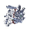
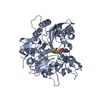
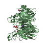
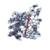
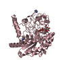
| Title | Crystal Structure of a Hypothetical Protein from Neisseria gonorrhoeae with bound ppGpp | ||||||
 Components Components | Putative phosphoribosyltransferase | ||||||
 Keywords Keywords | TRANSFERASE / SSGCID / ppGpp / possible Phosphoribosyltransferase / Structural Genomics / Seattle Structural Genomics Center for Infectious Disease | ||||||
| Function / homology | Rossmann fold - #2020 / Phosphoribosyl transferase domain / Phosphoribosyltransferase-like / Phosphoribosyltransferase domain / Rossmann fold / 3-Layer(aba) Sandwich / Alpha Beta / GUANOSINE-5',3'-TETRAPHOSPHATE / Phosphoribosyltransferase Function and homology information Function and homology information | ||||||
| Biological species |  Neisseria gonorrhoeae (bacteria) Neisseria gonorrhoeae (bacteria) | ||||||
| Method |  X-RAY DIFFRACTION / SYNCHROTRON / MOLECULAR REPLACEMENT / molecular replacement / Resolution: 1.5 Å X-RAY DIFFRACTION / SYNCHROTRON / MOLECULAR REPLACEMENT / molecular replacement / Resolution: 1.5 Å | ||||||
 Authors Authors | Seattle Structural Genomics Center for Infectious Disease (SSGCID) | ||||||
 Citation Citation | Journal: to be published Title: Crystal Structure of a Hypothetical Protein from Neisseria gonorrhoeae with bound ppGpp Authors: Dranow, D.M. / Abendroth, J. / Lorimer, D.D. / Edwards, T.E. | ||||||
| History |
|
- Structure visualization
| Structure viewer | Molecule: MolmilJmol/JSmol |
|---|
- Downloads & links
-Download
| PDBx/mmCIF format | 5vog.cif.gz | 99.1 KB | Display | PDBx/mmCIF format |
|---|---|---|---|---|
| PDB format | pdb5vog.ent.gz | 71.3 KB | Display | PDB format |
| PDBx/mmJSON format | 5vog.json.gz | Tree view | PDBx/mmJSON format | |
| Others |  Other downloads Other downloads |
-Validation report
| Arichive directory | https://data.pdbj.org/pub/pdb/validation_reports/vo/5vogftp://data.pdbj.org/pub/pdb/validation_reports/vo/5vog | HTTPS FTP |
|---|
-Related structure data
| Related structure data |  2jkzS S: Starting model for refinement |
|---|---|
| Similar structure data | |
| Other databases |


-Links
 PDBj
PDBj
- Assembly
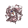
| Deposited unit | 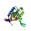
| |||||||||
|---|---|---|---|---|---|---|---|---|---|---|
| 1 | 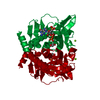
| |||||||||
| Unit cell |
| |||||||||
| Components on special symmetry positions |
|
-Components
| #1: Protein | Mass: 20977.008 Da / Num. of mol.: 1 Source method: isolated from a genetically manipulated source Source: (gene. exp.) Neisseria gonorrhoeae (bacteria)Gene: ESCNG_40048, WHOF_00431, WHOF_02526C, WHOG_01129, WHOG_02374C, WHOK_01250, WHOK_02372C, WHOM_00300, WHOM_02380C, WHON_00453, WHON_02375C, WHOU_01208, WHOU_02454C, WHOV_00719, WHOV_02442C, WHOW_ ...Gene: ESCNG_40048, WHOF_00431, WHOF_02526C, WHOG_01129, WHOG_02374C, WHOK_01250, WHOK_02372C, WHOM_00300, WHOM_02380C, WHON_00453, WHON_02375C, WHOU_01208, WHOU_02454C, WHOV_00719, WHOV_02442C, WHOW_01101, WHOW_02437C, WHOX_00398, WHOX_02372C, WHOZ_01227, WHOZ_02445C Plasmid: NegoA.19192.a.B1 / Production host: | ||||||
|---|---|---|---|---|---|---|---|
| #2: Chemical |   Mass: 62.068 Da / Num. of mol.: 2 / Source method: obtained synthetically / Formula: C2H6O2 Mass: 62.068 Da / Num. of mol.: 2 / Source method: obtained synthetically / Formula: C2H6O2#3: Chemical | ChemComp-G4P / | 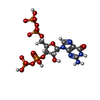  Type: RNA linking / Mass: 603.160 Da / Num. of mol.: 1 / Source method: obtained synthetically / Formula: C10H17N5O17P4 Type: RNA linking / Mass: 603.160 Da / Num. of mol.: 1 / Source method: obtained synthetically / Formula: C10H17N5O17P4#4: Chemical |   Mass: 24.305 Da / Num. of mol.: 2 / Source method: obtained synthetically / Formula: Mg Mass: 24.305 Da / Num. of mol.: 2 / Source method: obtained synthetically / Formula: Mg#5: Water | ChemComp-HOH / |  Mass: 18.015 Da / Num. of mol.: 201 / Source method: isolated from a natural source / Formula: H2O Mass: 18.015 Da / Num. of mol.: 201 / Source method: isolated from a natural source / Formula: H2O |
-Experimental details
-Experiment
| Experiment | Method: X-RAY DIFFRACTION / Number of used crystals: 1 |
|---|
- Sample preparation
| Crystal | Density Matthews: 2.66 Å3/Da / Density % sol: 53.69 % |
|---|---|
| Crystal grow | Temperature: 290 K / Method: vapor diffusion, sitting drop / pH: 5.5 Details: NegoA.19192.a.B1.PS38045 at 17 mg/ml mixed 1:1 with Morpheus(a2): 10% (w/v) PEG-8000, 20% (v/v) ethylene glycol, 0.1 M MES/imidazole, pH = 5.5, 0.03 M each magnesium chloride and calcium chloride, direct cryo |
-Data collection
| Diffraction | Mean temperature: 100 K | ||||||||||||||||||||||||||||||||||||||||||||||||||||||||||||||||||||||||||||||||||||||||||||||||||||||||||||||||||||||||||||||||||||||||||||||||||||||||||||||||||||||||
|---|---|---|---|---|---|---|---|---|---|---|---|---|---|---|---|---|---|---|---|---|---|---|---|---|---|---|---|---|---|---|---|---|---|---|---|---|---|---|---|---|---|---|---|---|---|---|---|---|---|---|---|---|---|---|---|---|---|---|---|---|---|---|---|---|---|---|---|---|---|---|---|---|---|---|---|---|---|---|---|---|---|---|---|---|---|---|---|---|---|---|---|---|---|---|---|---|---|---|---|---|---|---|---|---|---|---|---|---|---|---|---|---|---|---|---|---|---|---|---|---|---|---|---|---|---|---|---|---|---|---|---|---|---|---|---|---|---|---|---|---|---|---|---|---|---|---|---|---|---|---|---|---|---|---|---|---|---|---|---|---|---|---|---|---|---|---|---|---|---|
| Diffraction source | Source: SYNCHROTRON / Site: ALS  / Beamline: 5.0.2 / Wavelength: 1 Å / Beamline: 5.0.2 / Wavelength: 1 Å | ||||||||||||||||||||||||||||||||||||||||||||||||||||||||||||||||||||||||||||||||||||||||||||||||||||||||||||||||||||||||||||||||||||||||||||||||||||||||||||||||||||||||
| Detector | Type: DECTRIS PILATUS3 6M / Detector: PIXEL / Date: Apr 29, 2017 | ||||||||||||||||||||||||||||||||||||||||||||||||||||||||||||||||||||||||||||||||||||||||||||||||||||||||||||||||||||||||||||||||||||||||||||||||||||||||||||||||||||||||
| Radiation | Monochromator: Double-crystal, Si(111) / Protocol: SINGLE WAVELENGTH / Monochromatic (M) / Laue (L): M / Scattering type: x-ray | ||||||||||||||||||||||||||||||||||||||||||||||||||||||||||||||||||||||||||||||||||||||||||||||||||||||||||||||||||||||||||||||||||||||||||||||||||||||||||||||||||||||||
| Radiation wavelength | Wavelength: 1 Å / Relative weight: 1 | ||||||||||||||||||||||||||||||||||||||||||||||||||||||||||||||||||||||||||||||||||||||||||||||||||||||||||||||||||||||||||||||||||||||||||||||||||||||||||||||||||||||||
| Reflection | Resolution: 1.5→40.635 Å / Num. obs: 35700 / % possible obs: 99.7 % / Observed criterion σ(I): -3 / Redundancy: 5.493 % / Biso Wilson estimate: 24.57 Å2 / CC1/2: 0.999 / Rmerge(I) obs: 0.031 / Rrim(I) all: 0.034 / Χ2: 0.993 / Net I/σ(I): 26.34 | ||||||||||||||||||||||||||||||||||||||||||||||||||||||||||||||||||||||||||||||||||||||||||||||||||||||||||||||||||||||||||||||||||||||||||||||||||||||||||||||||||||||||
| Reflection shell | Diffraction-ID: 1
|
-Phasing
| Phasing | Method: molecular replacement |
|---|
- Processing
| Software |
| |||||||||||||||||||||||||||||||||||||||||||||||||||||||||||||||||||||||||||||||||||||||||||||||||||||||||
|---|---|---|---|---|---|---|---|---|---|---|---|---|---|---|---|---|---|---|---|---|---|---|---|---|---|---|---|---|---|---|---|---|---|---|---|---|---|---|---|---|---|---|---|---|---|---|---|---|---|---|---|---|---|---|---|---|---|---|---|---|---|---|---|---|---|---|---|---|---|---|---|---|---|---|---|---|---|---|---|---|---|---|---|---|---|---|---|---|---|---|---|---|---|---|---|---|---|---|---|---|---|---|---|---|---|---|
| Refinement | Method to determine structure: MOLECULAR REPLACEMENT Starting model: 2JKZ Resolution: 1.5→40.635 Å / SU ML: 0.12 / Cross valid method: FREE R-VALUE / σ(F): 1.35 / Phase error: 17.19
| |||||||||||||||||||||||||||||||||||||||||||||||||||||||||||||||||||||||||||||||||||||||||||||||||||||||||
| Solvent computation | Shrinkage radii: 0.9 Å / VDW probe radii: 1.11 Å | |||||||||||||||||||||||||||||||||||||||||||||||||||||||||||||||||||||||||||||||||||||||||||||||||||||||||
| Displacement parameters | Biso max: 113.28 Å2 / Biso mean: 35.5166 Å2 / Biso min: 18.86 Å2 | |||||||||||||||||||||||||||||||||||||||||||||||||||||||||||||||||||||||||||||||||||||||||||||||||||||||||
| Refinement step | Cycle: final / Resolution: 1.5→40.635 Å
| |||||||||||||||||||||||||||||||||||||||||||||||||||||||||||||||||||||||||||||||||||||||||||||||||||||||||
| Refine LS restraints |
| |||||||||||||||||||||||||||||||||||||||||||||||||||||||||||||||||||||||||||||||||||||||||||||||||||||||||
| LS refinement shell | Refine-ID: X-RAY DIFFRACTION / Rfactor Rfree error: 0 / Total num. of bins used: 14
|
