 Movie
Movie Controller

[English] 日本語
 Yorodumi
- PDB-6ehn: Structural insight into a promiscuous CE15 esterase from the mari... -
+ Open data

- Basic information
| Entry | Database: PDB / ID: 6ehn | |||||||||
|---|---|---|---|---|---|---|---|---|---|---|
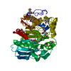
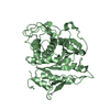
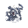
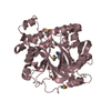
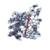
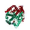
| Title | Structural insight into a promiscuous CE15 esterase from the marine bacterial metagenome | |||||||||
 Components Components | Carbohydrate esterase MZ0003 | |||||||||
 Keywords Keywords | HYDROLASE / esterase / protein structure | |||||||||
| Function / homology | Glucuronyl esterase, fungi / Hydrolases; Acting on ester bonds; Carboxylic-ester hydrolases / carboxylic ester hydrolase activity / polysaccharide catabolic process / Alpha/Beta hydrolase fold / periplasmic space / Carbohydrate esterase MZ0003 Function and homology information Function and homology information | |||||||||
| Biological species |  Unknown prokaryotic organism (environmental samples) Unknown prokaryotic organism (environmental samples) | |||||||||
| Method |  X-RAY DIFFRACTION / SYNCHROTRON / SAD / molecular replacement / Resolution: 1.9 Å X-RAY DIFFRACTION / SYNCHROTRON / SAD / molecular replacement / Resolution: 1.9 Å | |||||||||
 Authors Authors | Helland, R. / De Santi, C. / Gani, O. / Williamson, A.K. | |||||||||
| Funding support |  Norway, 2items Norway, 2items
| |||||||||
 Citation Citation | Journal: Sci Rep / Year: 2017 Title: Structural insight into a CE15 esterase from the marine bacterial metagenome. Authors: De Santi, C. / Gani, O.A. / Helland, R. / Williamson, A. | |||||||||
| History |
|
- Structure visualization
| Structure viewer | Molecule: MolmilJmol/JSmol |
|---|
- Downloads & links
-Download
| PDBx/mmCIF format | 6ehn.cif.gz | 100.1 KB | Display | PDBx/mmCIF format |
|---|---|---|---|---|
| PDB format | pdb6ehn.ent.gz | 74.4 KB | Display | PDB format |
| PDBx/mmJSON format | 6ehn.json.gz | Tree view | PDBx/mmJSON format | |
| Others |  Other downloads Other downloads |
-Validation report
| Arichive directory | https://data.pdbj.org/pub/pdb/validation_reports/eh/6ehnftp://data.pdbj.org/pub/pdb/validation_reports/eh/6ehn | HTTPS FTP |
|---|
-Related structure data
| Similar structure data |
|---|


-Links
 PDBj
PDBj- Assembly
| Deposited unit | 
| ||||||||
|---|---|---|---|---|---|---|---|---|---|
| 1 |
| ||||||||
| Unit cell |
|
-Components
| #1: Protein | Mass: 44889.824 Da / Num. of mol.: 1 Source method: isolated from a genetically manipulated source Source: (gene. exp.) Unknown prokaryotic organism (environmental samples)Gene: MZ0003 / Production host: References: UniProt: A0A0K2VM55, Hydrolases; Acting on ester bonds; Carboxylic-ester hydrolases |
|---|---|
| #2: Chemical | ChemComp-GOL /   Mass: 92.094 Da / Num. of mol.: 1 / Source method: obtained synthetically / Formula: C3H8O3 Mass: 92.094 Da / Num. of mol.: 1 / Source method: obtained synthetically / Formula: C3H8O3 |
| #3: Water | ChemComp-HOH /  Mass: 18.015 Da / Num. of mol.: 315 / Source method: isolated from a natural source / Formula: H2O Mass: 18.015 Da / Num. of mol.: 315 / Source method: isolated from a natural source / Formula: H2O |
-Experimental details
-Experiment
| Experiment | Method: X-RAY DIFFRACTION / Number of used crystals: 1 |
|---|
- Sample preparation
| Crystal | Density Matthews: 3.07 Å3/Da / Density % sol: 59.97 % |
|---|---|
| Crystal grow | Temperature: 277 K / Method: vapor diffusion, hanging drop / pH: 6 / Details: PEG 3350, 24% NaCitrate, pH 6, 0.1 M |
-Data collection
| Diffraction | Mean temperature: 100 K | ||||||||||||||||||||||||
|---|---|---|---|---|---|---|---|---|---|---|---|---|---|---|---|---|---|---|---|---|---|---|---|---|---|
| Diffraction source | Source: SYNCHROTRON / Site: BESSY  / Beamline: 14.1 / Wavelength: 0.91840, 1.28202 / Beamline: 14.1 / Wavelength: 0.91840, 1.28202 | ||||||||||||||||||||||||
| Detector | Type: MARMOSAIC 225 mm CCD / Detector: CCD / Date: Jan 24, 2015 | ||||||||||||||||||||||||
| Radiation | Protocol: SINGLE WAVELENGTH / Monochromatic (M) / Laue (L): M / Scattering type: x-ray | ||||||||||||||||||||||||
| Radiation wavelength |
| ||||||||||||||||||||||||
| Reflection | Resolution: 1.9→47 Å / Num. obs: 43689 / % possible obs: 99.9 % / Redundancy: 5.6 % / Biso Wilson estimate: 25.48 Å2 / CC1/2: 0.999 / Rmerge(I) obs: 0.067 / Rpim(I) all: 0.031 / Rrim(I) all: 0.074 / Net I/av σ(I): 16.4 / Net I/σ(I): 16.4 | ||||||||||||||||||||||||
| Reflection shell | Diffraction-ID: 1
|
-Phasing
| Phasing |
|
|---|
- Processing
| Software |
| ||||||||||||||||||||||||||||||||||||||||||||||||||||||||||||
|---|---|---|---|---|---|---|---|---|---|---|---|---|---|---|---|---|---|---|---|---|---|---|---|---|---|---|---|---|---|---|---|---|---|---|---|---|---|---|---|---|---|---|---|---|---|---|---|---|---|---|---|---|---|---|---|---|---|---|---|---|---|
| Refinement | Method to determine structure: SAD / Resolution: 1.9→47 Å / Cor.coef. Fo:Fc: 0.962 / Cor.coef. Fo:Fc free: 0.944 / SU B: 3.427 / SU ML: 0.097 / Cross valid method: THROUGHOUT / σ(F): 0 / ESU R: 0.125 / ESU R Free: 0.125 Details: HYDROGENS HAVE BEEN ADDED IN THE RIDING POSITIONS U VALUES : REFINED INDIVIDUALLY
| ||||||||||||||||||||||||||||||||||||||||||||||||||||||||||||
| Solvent computation | Ion probe radii: 0.8 Å / Shrinkage radii: 0.8 Å / VDW probe radii: 1.2 Å | ||||||||||||||||||||||||||||||||||||||||||||||||||||||||||||
| Displacement parameters | Biso max: 122.43 Å2 / Biso mean: 33.169 Å2 / Biso min: 7.62 Å2
| ||||||||||||||||||||||||||||||||||||||||||||||||||||||||||||
| Refinement step | Cycle: final / Resolution: 1.9→47 Å
| ||||||||||||||||||||||||||||||||||||||||||||||||||||||||||||
| Refine LS restraints |
| ||||||||||||||||||||||||||||||||||||||||||||||||||||||||||||
| LS refinement shell | Resolution: 1.9→1.949 Å / Rfactor Rfree error: 0 / Total num. of bins used: 20
|
