 Movie
Movie Controller

[English] 日本語
 Yorodumi
- PDB-5vmp: Crystal Structure of Human KDM4 with Small Molecule Inhibitor QC5714 -
+ Open data

- Basic information
| Entry | Database: PDB / ID: 5vmp | ||||||
|---|---|---|---|---|---|---|---|
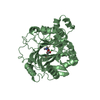
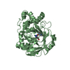
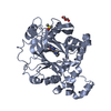
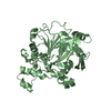
| Title | Crystal Structure of Human KDM4 with Small Molecule Inhibitor QC5714 | ||||||
 Components Components | Lysine-specific demethylase 4A | ||||||
 Keywords Keywords | OXIDOREDUCTASE/INHIBITOR / KDM4 / Inhibitor-complex / demethylase / epigenetics / OXIDOREDUCTASE-INHIBITOR complex | ||||||
| Function / homology |  Function and homology information Function and homology information[histone H3]-trimethyl-L-lysine36 demethylase / histone H3K36me2/H3K36me3 demethylase activity / histone H4K20me2 reader activity / histone H3K36 demethylase activity / [histone H3]-trimethyl-L-lysine9 demethylase / histone H3K9me2/H3K9me3 demethylase activity / histone H3K9 demethylase activity / histone demethylase activity / pericentric heterochromatin / NR1H3 & NR1H2 regulate gene expression linked to cholesterol transport and efflux ...[histone H3]-trimethyl-L-lysine36 demethylase / histone H3K36me2/H3K36me3 demethylase activity / histone H4K20me2 reader activity / histone H3K36 demethylase activity / [histone H3]-trimethyl-L-lysine9 demethylase / histone H3K9me2/H3K9me3 demethylase activity / histone H3K9 demethylase activity / histone demethylase activity / pericentric heterochromatin / NR1H3 & NR1H2 regulate gene expression linked to cholesterol transport and efflux / negative regulation of autophagy / HDMs demethylate histones / fibrillar center / Recruitment and ATM-mediated phosphorylation of repair and signaling proteins at DNA double strand breaks / regulation of gene expression / chromatin remodeling / negative regulation of gene expression / negative regulation of DNA-templated transcription / ubiquitin protein ligase binding / chromatin / DNA-templated transcription / zinc ion binding / nucleoplasm / nucleus Similarity search - Function | ||||||
| Biological species |  Homo sapiens (human) Homo sapiens (human) | ||||||
| Method |  X-RAY DIFFRACTION / SYNCHROTRON / MOLECULAR REPLACEMENT / Resolution: 2.48 Å X-RAY DIFFRACTION / SYNCHROTRON / MOLECULAR REPLACEMENT / Resolution: 2.48 Å | ||||||
 Authors Authors | Hosfield, D.J. | ||||||
 Citation Citation | Journal: ACS Med Chem Lett / Year: 2017 Title: Design of KDM4 Inhibitors with Antiproliferative Effects in Cancer Models. Authors: Chen, Y.K. / Bonaldi, T. / Cuomo, A. / Del Rosario, J.R. / Hosfield, D.J. / Kanouni, T. / Kao, S.C. / Lai, C. / Lobo, N.A. / Matuszkiewicz, J. / McGeehan, A. / O'Connell, S.M. / Shi, L. / ...Authors: Chen, Y.K. / Bonaldi, T. / Cuomo, A. / Del Rosario, J.R. / Hosfield, D.J. / Kanouni, T. / Kao, S.C. / Lai, C. / Lobo, N.A. / Matuszkiewicz, J. / McGeehan, A. / O'Connell, S.M. / Shi, L. / Stafford, J.A. / Stansfield, R.K. / Veal, J.M. / Weiss, M.S. / Yuen, N.Y. / Wallace, M.B. | ||||||
| History |
|
- Structure visualization
| Structure viewer | Molecule: MolmilJmol/JSmol |
|---|
- Downloads & links
-Download
| PDBx/mmCIF format | 5vmp.cif.gz | 296 KB | Display | PDBx/mmCIF format |
|---|---|---|---|---|
| PDB format | pdb5vmp.ent.gz | 239.1 KB | Display | PDB format |
| PDBx/mmJSON format | 5vmp.json.gz | Tree view | PDBx/mmJSON format | |
| Others |  Other downloads Other downloads |
-Validation report
| Arichive directory | https://data.pdbj.org/pub/pdb/validation_reports/vm/5vmpftp://data.pdbj.org/pub/pdb/validation_reports/vm/5vmp | HTTPS FTP |
|---|
-Related structure data
| Related structure data |  5vgiC  3pdqS S: Starting model for refinement C: citing same article ( |
|---|---|
| Similar structure data |

-Links
 PDBj
PDBj


- Assembly
| Deposited unit | 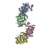
| ||||||||
|---|---|---|---|---|---|---|---|---|---|
| 1 | 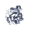
| ||||||||
| 2 | 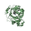
| ||||||||
| 3 | 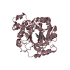
| ||||||||
| 4 | 
| ||||||||
| Unit cell |
|
-Components
| #1: Protein | Mass: 42975.770 Da / Num. of mol.: 4 Source method: isolated from a genetically manipulated source Source: (gene. exp.) Homo sapiens (human) / Gene: KDM4A, JHDM3A, JMJD2, JMJD2A, KIAA0677 / Production host:  References: UniProt: O75164, Oxidoreductases; Acting on paired donors, with incorporation or reduction of molecular oxygen; With 2-oxoglutarate as one donor, and incorporation of one atom of oxygen into each donor #2: Chemical | ChemComp-NI /   Mass: 58.693 Da / Num. of mol.: 4 / Source method: obtained synthetically / Formula: Ni Mass: 58.693 Da / Num. of mol.: 4 / Source method: obtained synthetically / Formula: Ni#3: Chemical | ChemComp-ZN /   Mass: 65.409 Da / Num. of mol.: 4 / Source method: obtained synthetically / Formula: Zn Mass: 65.409 Da / Num. of mol.: 4 / Source method: obtained synthetically / Formula: Zn#4: Chemical | ChemComp-9FJ /   Mass: 312.363 Da / Num. of mol.: 4 / Source method: obtained synthetically / Formula: C18H20N2O3 Mass: 312.363 Da / Num. of mol.: 4 / Source method: obtained synthetically / Formula: C18H20N2O3#5: Water | ChemComp-HOH / |  Mass: 18.015 Da / Num. of mol.: 244 / Source method: isolated from a natural source / Formula: H2O Mass: 18.015 Da / Num. of mol.: 244 / Source method: isolated from a natural source / Formula: H2O |
|---|
-Experimental details
-Experiment
| Experiment | Method: X-RAY DIFFRACTION / Number of used crystals: 1 |
|---|
- Sample preparation
| Crystal | Density Matthews: 2.57 Å3/Da / Density % sol: 52.07 % |
|---|---|
| Crystal grow | Temperature: 293 K / Method: vapor diffusion / Details: 22% PEG4K, 100mM HEPES pH 7.5 |
-Data collection
| Diffraction | Mean temperature: 77 K | |||||||||||||||||||||||||||||||||||||||||||||||||||||||||||||||||||||||||||||
|---|---|---|---|---|---|---|---|---|---|---|---|---|---|---|---|---|---|---|---|---|---|---|---|---|---|---|---|---|---|---|---|---|---|---|---|---|---|---|---|---|---|---|---|---|---|---|---|---|---|---|---|---|---|---|---|---|---|---|---|---|---|---|---|---|---|---|---|---|---|---|---|---|---|---|---|---|---|---|
| Diffraction source | Source: SYNCHROTRON / Site: APS  / Beamline: 17-ID / Wavelength: 1 Å / Beamline: 17-ID / Wavelength: 1 Å | |||||||||||||||||||||||||||||||||||||||||||||||||||||||||||||||||||||||||||||
| Detector | Type: ADSC QUANTUM 210 / Detector: CCD / Date: Jun 25, 2016 | |||||||||||||||||||||||||||||||||||||||||||||||||||||||||||||||||||||||||||||
| Radiation | Protocol: SINGLE WAVELENGTH / Monochromatic (M) / Laue (L): M / Scattering type: x-ray | |||||||||||||||||||||||||||||||||||||||||||||||||||||||||||||||||||||||||||||
| Radiation wavelength | Wavelength: 1 Å / Relative weight: 1 | |||||||||||||||||||||||||||||||||||||||||||||||||||||||||||||||||||||||||||||
| Reflection | Resolution: 2.48→50 Å / Num. obs: 57177 / % possible obs: 99 % / Redundancy: 3.7 % / Rmerge(I) obs: 0.127 / Χ2: 1.035 / Net I/σ(I): 5.7 / Num. measured all: 214006 | |||||||||||||||||||||||||||||||||||||||||||||||||||||||||||||||||||||||||||||
| Reflection shell |
|
- Processing
| Software |
| ||||||||||||||||||||||||||||||||||||||||||||||||||||||||||||
|---|---|---|---|---|---|---|---|---|---|---|---|---|---|---|---|---|---|---|---|---|---|---|---|---|---|---|---|---|---|---|---|---|---|---|---|---|---|---|---|---|---|---|---|---|---|---|---|---|---|---|---|---|---|---|---|---|---|---|---|---|---|
| Refinement | Method to determine structure: MOLECULAR REPLACEMENT Starting model: 3PDQ Resolution: 2.48→50 Å / Cor.coef. Fo:Fc: 0.944 / Cor.coef. Fo:Fc free: 0.902 / WRfactor Rfree: 0.2598 / WRfactor Rwork: 0.1914 / FOM work R set: 0.7918 / SU B: 10.99 / SU ML: 0.239 / SU R Cruickshank DPI: 0.5994 / SU Rfree: 0.305 / Cross valid method: THROUGHOUT / σ(F): 0 / ESU R: 0.599 / ESU R Free: 0.305 / Stereochemistry target values: MAXIMUM LIKELIHOOD Details: HYDROGENS HAVE BEEN ADDED IN THE RIDING POSITIONS U VALUES : REFINED INDIVIDUALLY
| ||||||||||||||||||||||||||||||||||||||||||||||||||||||||||||
| Solvent computation | Ion probe radii: 0.8 Å / Shrinkage radii: 0.8 Å / VDW probe radii: 1.2 Å / Solvent model: MASK | ||||||||||||||||||||||||||||||||||||||||||||||||||||||||||||
| Displacement parameters | Biso max: 121.86 Å2 / Biso mean: 41.597 Å2 / Biso min: 13.69 Å2
| ||||||||||||||||||||||||||||||||||||||||||||||||||||||||||||
| Refinement step | Cycle: final / Resolution: 2.48→50 Å
| ||||||||||||||||||||||||||||||||||||||||||||||||||||||||||||
| Refine LS restraints |
| ||||||||||||||||||||||||||||||||||||||||||||||||||||||||||||
| LS refinement shell | Resolution: 2.481→2.545 Å / Rfactor Rfree error: 0 / Total num. of bins used: 20
|
