negative regulation of non-canonical inflammasome complex assembly / protein N-acetylglucosaminyltransferase complex / regulation of insulin receptor signaling pathway / protein O-acetylglucosaminyltransferase activity / protein O-GlcNAc transferase / positive regulation of transcription from RNA polymerase II promoter by glucose / acetylglucosaminyltransferase activity / regulation of Rac protein signal transduction / regulation of necroptotic process / negative regulation of stem cell population maintenance ...negative regulation of non-canonical inflammasome complex assembly / protein N-acetylglucosaminyltransferase complex / regulation of insulin receptor signaling pathway / protein O-acetylglucosaminyltransferase activity / protein O-GlcNAc transferase / positive regulation of transcription from RNA polymerase II promoter by glucose / acetylglucosaminyltransferase activity / regulation of Rac protein signal transduction / regulation of necroptotic process / negative regulation of stem cell population maintenance / protein O-linked glycosylation / NSL complex / regulation of chromosome separation / positive regulation of aggrephagy / WNT mediated activation of DVL / Condensation of Prometaphase Chromosomes / protein kinase CK2 complex / symbiont-mediated disruption of host cell PML body / regulation of glycolytic process / RIPK1-mediated regulated necrosis / regulation of gluconeogenesis / Receptor Mediated Mitophagy / regulation of synapse assembly / Formation of WDR5-containing histone-modifying complexes / Synthesis of PC / Sin3-type complex / regulation of neurotransmitter receptor localization to postsynaptic specialization membrane / RUNX1 interacts with co-factors whose precise effect on RUNX1 targets is not known / negative regulation of signal transduction by p53 class mediator / Maturation of hRSV A proteins / positive regulation of stem cell population maintenance / phosphatidylinositol-3,4,5-trisphosphate binding / hemopoiesis / positive regulation of proteolysis / negative regulation of apoptotic signaling pathway / histone acetyltransferase complex / positive regulation of Wnt signaling pathway / negative regulation of double-strand break repair via homologous recombination / positive regulation of lipid biosynthetic process / mitophagy / : / negative regulation of proteasomal ubiquitin-dependent protein catabolic process / response to nutrient / negative regulation of protein ubiquitination / positive regulation of TORC1 signaling / negative regulation of cell migration / positive regulation of translation / Signal transduction by L1 / cell projection / cellular response to glucose stimulus / circadian regulation of gene expression / Hsp90 protein binding / negative regulation of transforming growth factor beta receptor signaling pathway / response to insulin / PML body / protein processing / chromatin DNA binding / Regulation of necroptotic cell death / mitochondrial membrane / Regulation of PTEN stability and activity / Wnt signaling pathway / positive regulation of protein catabolic process / kinase activity / KEAP1-NFE2L2 pathway / UCH proteinases / rhythmic process / Cooperation of PDCL (PhLP1) and TRiC/CCT in G-protein beta folding / double-strand break repair / positive regulation of cold-induced thermogenesis / HATs acetylate histones / chromatin organization / positive regulation of cell growth / Regulation of TP53 Activity through Phosphorylation / non-specific serine/threonine protein kinase / regulation of cell cycle / negative regulation of translation / protein stabilization / protein serine kinase activity / protein serine/threonine kinase activity / positive regulation of cell population proliferation / apoptotic process / DNA damage response / regulation of transcription by RNA polymerase II / positive regulation of DNA-templated transcription / glutamatergic synapse / negative regulation of transcription by RNA polymerase II / signal transduction / positive regulation of transcription by RNA polymerase II / protein-containing complex / nucleoplasm / ATP binding / identical protein binding / nucleus / plasma membrane / cytosol Similarity search - Function
Signal recognition particle alu RNA binding heterodimer, srp9/1 - #150 / UDP-N-acetylglucosamine--peptide N-acetylglucosaminyltransferase 110kDa subunit / Rossmann fold - #11380 / O-GlcNAc transferase, C-terminal / Glycosyl transferase family 41 / Signal recognition particle alu RNA binding heterodimer, srp9/1 / TPR repeat / Tetratricopeptide repeat / Casein Kinase 2, subunit alpha / Tetratricopeptide repeat ...Signal recognition particle alu RNA binding heterodimer, srp9/1 - #150 / UDP-N-acetylglucosamine--peptide N-acetylglucosaminyltransferase 110kDa subunit / Rossmann fold - #11380 / O-GlcNAc transferase, C-terminal / Glycosyl transferase family 41 / Signal recognition particle alu RNA binding heterodimer, srp9/1 / TPR repeat / Tetratricopeptide repeat / Casein Kinase 2, subunit alpha / Tetratricopeptide repeat / Tetratricopeptide repeat / Glycogen Phosphorylase B; / TPR repeat region circular profile. / TPR repeat profile. / Tetratricopeptide repeats / Tetratricopeptide repeat / Tetratricopeptide-like helical domain superfamily / Serine/threonine-protein kinase, active site / Serine/Threonine protein kinases active-site signature. / Protein kinase domain / Serine/Threonine protein kinases, catalytic domain / Protein kinase, ATP binding site / Protein kinases ATP-binding region signature. / Protein kinase domain profile. / Protein kinase domain / Protein kinase-like domain superfamily / Rossmann fold / 2-Layer Sandwich / 3-Layer(aba) Sandwich / Alpha Beta Similarity search - Domain/homology
 Movie
Movie Controller

 Yorodumi
 Open data
 Basic information
 Components
 Keywords
 Function and homology information
 Homo sapiens (human)
 X-RAY DIFFRACTION /
  Authors
 United States, 1items
  Citation
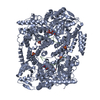
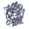
 Structure visualization
 Downloads & links
 Other downloads


 PDBj
PDBj


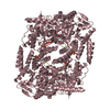
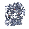
 Assembly
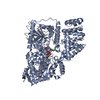


 Type: RNA linking / Mass: 404.161 Da / Num. of mol.: 1 / Source method: obtained synthetically / Formula: C9H14N2O12P2 / Comment: UDP*YM
 Type: D-saccharide, beta linking / Mass: 281.690 Da / Num. of mol.: 1 / Source method: obtained synthetically / Formula: C10H16ClNO6
 Mass: 18.015 Da / Num. of mol.: 191 / Source method: isolated from a natural source / Formula: H2O
 Sample preparation
 Processing