negative regulation of non-canonical inflammasome complex assembly / protein N-acetylglucosaminyltransferase complex / regulation of insulin receptor signaling pathway / protein O-GlcNAc transferase / protein O-acetylglucosaminyltransferase activity / positive regulation of transcription from RNA polymerase II promoter by glucose / acetylglucosaminyltransferase activity / regulation of Rac protein signal transduction / regulation of necroptotic process / negative regulation of stem cell population maintenance ...negative regulation of non-canonical inflammasome complex assembly / protein N-acetylglucosaminyltransferase complex / regulation of insulin receptor signaling pathway / protein O-GlcNAc transferase / protein O-acetylglucosaminyltransferase activity / positive regulation of transcription from RNA polymerase II promoter by glucose / acetylglucosaminyltransferase activity / regulation of Rac protein signal transduction / regulation of necroptotic process / negative regulation of stem cell population maintenance / protein O-linked glycosylation / Phosphorylation and nuclear translocation of BMAL1 (ARNTL) and CLOCK / NSL complex / positive regulation of aggrephagy / regulation of chromosome separation / WNT mediated activation of DVL / Condensation of Prometaphase Chromosomes / regulation of glycolytic process / protein kinase CK2 complex / symbiont-mediated disruption of host cell PML body / regulation of gluconeogenesis / RIPK1-mediated regulated necrosis / Phosphorylation and nuclear translocation of the CRY:PER:kinase complex / Regulation of CDH1 posttranslational processing and trafficking to plasma membrane / Receptor Mediated Mitophagy / Formation of WDR5-containing histone-modifying complexes / regulation of synapse assembly / Synthesis of PC / Sin3-type complex / positive regulation of stem cell population maintenance / regulation of neurotransmitter receptor localization to postsynaptic specialization membrane / positive regulation of proteolysis / negative regulation of signal transduction by p53 class mediator / phosphatidylinositol-3,4,5-trisphosphate binding / RUNX1 interacts with co-factors whose precise effect on RUNX1 targets is not known / Maturation of hRSV A proteins / hemopoiesis / histone acetyltransferase complex / negative regulation of apoptotic signaling pathway / negative regulation of double-strand break repair via homologous recombination / positive regulation of lipid biosynthetic process / positive regulation of Wnt signaling pathway / mitophagy / cell projection / response to nutrient / positive regulation of TORC1 signaling / negative regulation of protein ubiquitination / negative regulation of proteasomal ubiquitin-dependent protein catabolic process / negative regulation of cell migration / Signal transduction by L1 / positive regulation of translation / circadian regulation of gene expression / negative regulation of transforming growth factor beta receptor signaling pathway / cellular response to glucose stimulus / protein processing / Hsp90 protein binding / chromatin DNA binding / PML body / response to insulin / Regulation of necroptotic cell death / mitochondrial membrane / Wnt signaling pathway / Regulation of PTEN stability and activity / kinase activity / positive regulation of protein catabolic process / UCH proteinases / KEAP1-NFE2L2 pathway / rhythmic process / double-strand break repair / Cooperation of PDCL (PhLP1) and TRiC/CCT in G-protein beta folding / positive regulation of cold-induced thermogenesis / HATs acetylate histones / chromatin organization / protein folding / positive regulation of cell growth / Regulation of TP53 Activity through Phosphorylation / non-specific serine/threonine protein kinase / regulation of cell cycle / negative regulation of translation / protein stabilization / protein serine kinase activity / protein serine/threonine kinase activity / apoptotic process / positive regulation of cell population proliferation / DNA damage response / regulation of transcription by RNA polymerase II / positive regulation of DNA-templated transcription / glutamatergic synapse / negative regulation of transcription by RNA polymerase II / signal transduction / positive regulation of transcription by RNA polymerase II / protein-containing complex / nucleoplasm / ATP binding / identical protein binding / nucleus / plasma membrane / cytosol Similarity search - Function
Signal recognition particle alu RNA binding heterodimer, srp9/1 - #150 / UDP-N-acetylglucosamine--peptide N-acetylglucosaminyltransferase 110kDa subunit / Rossmann fold - #11380 / O-GlcNAc transferase, C-terminal / Glycosyl transferase family 41 / Signal recognition particle alu RNA binding heterodimer, srp9/1 / TPR repeat / Tetratricopeptide repeat / Tetratricopeptide repeat / Casein Kinase 2, subunit alpha ...Signal recognition particle alu RNA binding heterodimer, srp9/1 - #150 / UDP-N-acetylglucosamine--peptide N-acetylglucosaminyltransferase 110kDa subunit / Rossmann fold - #11380 / O-GlcNAc transferase, C-terminal / Glycosyl transferase family 41 / Signal recognition particle alu RNA binding heterodimer, srp9/1 / TPR repeat / Tetratricopeptide repeat / Tetratricopeptide repeat / Casein Kinase 2, subunit alpha / Tetratricopeptide repeat / Glycogen Phosphorylase B; / TPR repeat region circular profile. / TPR repeat profile. / Tetratricopeptide repeats / Tetratricopeptide repeat / Tetratricopeptide-like helical domain superfamily / Serine/threonine-protein kinase, active site / Serine/Threonine protein kinases active-site signature. / Protein kinase domain / Serine/Threonine protein kinases, catalytic domain / Protein kinase, ATP binding site / Protein kinases ATP-binding region signature. / Protein kinase domain profile. / Protein kinase domain / Protein kinase-like domain superfamily / Rossmann fold / 2-Layer Sandwich / 3-Layer(aba) Sandwich / Alpha Beta Similarity search - Domain/homology
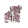
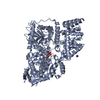
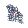
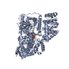
Mass: 80974.508 Da / Num. of mol.: 1 Source method: isolated from a genetically manipulated source Source: (gene. exp.) Homo sapiens (human) / Gene: OGT / Production host: Escherichia coli (E. coli) / References: UniProt: O15294, protein O-GlcNAc transferase
#2: Protein/peptide
TYR-PRO-GLY-GLY-SER-THR-PRO-VAL-SER-SER-ALA-ASN
Mass: 1398.562 Da / Num. of mol.: 1 / Source method: obtained synthetically / Source: (synth.) Homo sapiens (human) / References: UniProt: P68400*PLUS
In the structure databanks used in Yorodumi, some data are registered as the other names, "COVID-19 virus" and "2019-nCoV". Here are the details of the virus and the list of structure data.
Jan 31, 2019. EMDB accession codes are about to change! (news from PDBe EMDB page)
EMDB accession codes are about to change! (news from PDBe EMDB page)
The allocation of 4 digits for EMDB accession codes will soon come to an end. Whilst these codes will remain in use, new EMDB accession codes will include an additional digit and will expand incrementally as the available range of codes is exhausted. The current 4-digit format prefixed with “EMD-” (i.e. EMD-XXXX) will advance to a 5-digit format (i.e. EMD-XXXXX), and so on. It is currently estimated that the 4-digit codes will be depleted around Spring 2019, at which point the 5-digit format will come into force.
The EM Navigator/Yorodumi systems omit the EMD- prefix.
Related info.:Q: What is EMD? / ID/Accession-code notation in Yorodumi/EM Navigator
Yorodumi is a browser for structure data from EMDB, PDB, SASBDB, etc.
This page is also the successor to EM Navigator detail page, and also detail information page/front-end page for Omokage search.
The word "yorodu" (or yorozu) is an old Japanese word meaning "ten thousand". "mi" (miru) is to see.
Related info.:EMDB / PDB / SASBDB / Comparison of 3 databanks / Yorodumi Search / Aug 31, 2016. New EM Navigator & Yorodumi / Yorodumi Papers / Jmol/JSmol / Function and homology information / Changes in new EM Navigator and Yorodumi
 Movie
Movie Controller


 Open data
 Basic information
 Components
 Keywords
 Function and homology information
 Homo sapiens (human)
 X-RAY DIFFRACTION /
  Authors
 United States, 1items
  Citation
 Structure visualization
 Downloads & links
 Other downloads


 PDBj
PDBj


 Assembly
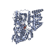


 Mass: 649.457 Da / Num. of mol.: 1 / Source method: obtained synthetically / Formula: C19H29N3O16P2S
 Mass: 18.015 Da / Num. of mol.: 191 / Source method: isolated from a natural source / Formula: H2O
 Sample preparation
 Processing