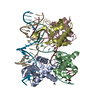
 Movie
Movie Controller


+ Open data

- Basic information
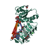
| Entry | Database: PDB / ID: 5vfy | ||||||
|---|---|---|---|---|---|---|---|
| Title | Structure of an accessory protein of the pCW3 relaxosome | ||||||
 Components Components | TcpK | ||||||
 Keywords Keywords | DNA BINDING PROTEIN / conjugation / winged helix-turn-helix / clostridium perfringens | ||||||
| Function / homology | Cch helix turn helix domain / Uncharacterized protein Function and homology information Function and homology information | ||||||
| Biological species |   Clostridium perfringens (bacteria) Clostridium perfringens (bacteria) | ||||||
| Method |  X-RAY DIFFRACTION / SYNCHROTRON / SAD / Resolution: 2.49 Å X-RAY DIFFRACTION / SYNCHROTRON / SAD / Resolution: 2.49 Å | ||||||
 Authors Authors | Traore, D.A.K. / Wisniewski, J.A. / Flanigan, S.F. / Conroy, P.J. / Panjikar, S. / Mok, Y.-F. / Lao, C. / Griffin, M.D.W. / Adams, V. / Rood, J.I. / Whisstock, J.C. | ||||||
 Citation Citation | Journal: Nat Commun / Year: 2018 Title: Crystal structure of TcpK in complex with oriT DNA of the antibiotic resistance plasmid pCW3. Authors: Traore, D.A.K. / Wisniewski, J.A. / Flanigan, S.F. / Conroy, P.J. / Panjikar, S. / Mok, Y.F. / Lao, C. / Griffin, M.D.W. / Adams, V. / Rood, J.I. / Whisstock, J.C. | ||||||
| History |
|
- Structure visualization
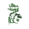
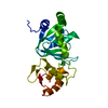
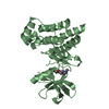
| Structure viewer | Molecule: MolmilJmol/JSmol |
|---|
- Downloads & links
-Download
| PDBx/mmCIF format | 5vfy.cif.gz | 100.3 KB | Display | PDBx/mmCIF format |
|---|---|---|---|---|
| PDB format | pdb5vfy.ent.gz | 77.6 KB | Display | PDB format |
| PDBx/mmJSON format | 5vfy.json.gz | Tree view | PDBx/mmJSON format | |
| Others |  Other downloads Other downloads |
-Validation report
| Arichive directory | https://data.pdbj.org/pub/pdb/validation_reports/vf/5vfyftp://data.pdbj.org/pub/pdb/validation_reports/vf/5vfy | HTTPS FTP |
|---|
-Related structure data


-Links
 PDBj
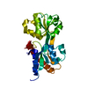
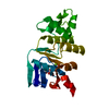
PDBj- Assembly
| Deposited unit | 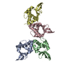
| ||||||||
|---|---|---|---|---|---|---|---|---|---|
| 1 | 
| ||||||||
| 2 | 
| ||||||||
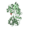
| Unit cell |
|
-Components
| #1: Protein | Mass: 13005.041 Da / Num. of mol.: 4 Source method: isolated from a genetically manipulated source Source: (gene. exp.) Clostridium perfringens (bacteria)Gene: ERS852446_03033, pBeta2_00022, pCpb2-CP1_20, pCW3_0028, pNetB-NE10_20, pNetB_00021, pTet_021 Plasmid: pGL12 / Production host: #2: Water | ChemComp-HOH / |  Mass: 18.015 Da / Num. of mol.: 187 / Source method: isolated from a natural source / Formula: H2O Mass: 18.015 Da / Num. of mol.: 187 / Source method: isolated from a natural source / Formula: H2O |
|---|
-Experimental details
-Experiment
| Experiment | Method: X-RAY DIFFRACTION / Number of used crystals: 1 |
|---|
- Sample preparation
| Crystal | Density Matthews: 2.5 Å3/Da / Density % sol: 50.72 % / Mosaicity: 0.29 ° |
|---|---|
| Crystal grow | Temperature: 293 K / Method: vapor diffusion, hanging drop / pH: 6.5 / Details: 1 M Na Citrate, 0.1 M Na Cacodylate, 0.1M Phenol |
-Data collection
| Diffraction | Mean temperature: 100 K | |||||||||||||||||||||
|---|---|---|---|---|---|---|---|---|---|---|---|---|---|---|---|---|---|---|---|---|---|---|
| Diffraction source | Source: SYNCHROTRON / Site: Australian Synchrotron  / Beamline: MX2 / Wavelength: 0.9537 Å / Beamline: MX2 / Wavelength: 0.9537 Å | |||||||||||||||||||||
| Detector | Type: ADSC QUANTUM 315r / Detector: CCD / Date: Mar 1, 2014 | |||||||||||||||||||||
| Radiation | Protocol: SINGLE WAVELENGTH / Monochromatic (M) / Laue (L): M / Scattering type: x-ray | |||||||||||||||||||||
| Radiation wavelength | Wavelength: 0.9537 Å / Relative weight: 1 | |||||||||||||||||||||
| Reflection | Resolution: 2.49→48.44 Å / Num. obs: 18698 / % possible obs: 99.5 % / Redundancy: 6.3 % / Biso Wilson estimate: 29.75 Å2 / CC1/2: 0.984 / Rmerge(I) obs: 0.214 / Rpim(I) all: 0.088 / Rrim(I) all: 0.232 / Net I/σ(I): 10.8 | |||||||||||||||||||||
| Reflection shell | Diffraction-ID: 1
|
- Processing
| Software |
| ||||||||||||||||||||||||||||||||||||||||||||||||||||||||||||||||||||||||||||||||||||||||||||||||||||||||||||
|---|---|---|---|---|---|---|---|---|---|---|---|---|---|---|---|---|---|---|---|---|---|---|---|---|---|---|---|---|---|---|---|---|---|---|---|---|---|---|---|---|---|---|---|---|---|---|---|---|---|---|---|---|---|---|---|---|---|---|---|---|---|---|---|---|---|---|---|---|---|---|---|---|---|---|---|---|---|---|---|---|---|---|---|---|---|---|---|---|---|---|---|---|---|---|---|---|---|---|---|---|---|---|---|---|---|---|---|---|---|
| Refinement | Method to determine structure: SAD / Resolution: 2.49→44.21 Å / Cor.coef. Fo:Fc: 0.904 / Cor.coef. Fo:Fc free: 0.855 / Rfactor Rfree error: 0 / SU R Cruickshank DPI: 0.42 / Cross valid method: THROUGHOUT / σ(F): 0 / SU R Blow DPI: 0.485 / SU Rfree Blow DPI: 0.259 / SU Rfree Cruickshank DPI: 0.255
| ||||||||||||||||||||||||||||||||||||||||||||||||||||||||||||||||||||||||||||||||||||||||||||||||||||||||||||
| Displacement parameters | Biso max: 123.97 Å2 / Biso mean: 27.58 Å2 / Biso min: 3 Å2
| ||||||||||||||||||||||||||||||||||||||||||||||||||||||||||||||||||||||||||||||||||||||||||||||||||||||||||||
| Refine analyze | Luzzati coordinate error obs: 0.29 Å | ||||||||||||||||||||||||||||||||||||||||||||||||||||||||||||||||||||||||||||||||||||||||||||||||||||||||||||
| Refinement step | Cycle: final / Resolution: 2.49→44.21 Å
| ||||||||||||||||||||||||||||||||||||||||||||||||||||||||||||||||||||||||||||||||||||||||||||||||||||||||||||
| Refine LS restraints |
| ||||||||||||||||||||||||||||||||||||||||||||||||||||||||||||||||||||||||||||||||||||||||||||||||||||||||||||
| LS refinement shell | Resolution: 2.49→2.64 Å / Rfactor Rfree error: 0 / Total num. of bins used: 9
|
