 Movie
Movie Controller


+ Open data

- Basic information
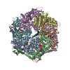
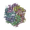
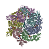
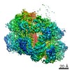
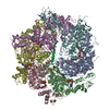
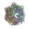
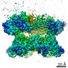
| Entry | Database: PDB / ID: 5vc7 | |||||||||
|---|---|---|---|---|---|---|---|---|---|---|
| Title | VCP like ATPase from T. acidophilum (VAT) - conformation 1 | |||||||||
 Components Components | VCP-like ATPase | |||||||||
 Keywords Keywords | HYDROLASE / AAA+ / ATPase / unfoldase | |||||||||
| Function / homology |  Function and homology information Function and homology information | |||||||||
| Biological species |   Thermoplasma acidophilum (acidophilic) Thermoplasma acidophilum (acidophilic) | |||||||||
| Method | ELECTRON MICROSCOPY / single particle reconstruction / cryo EM / Resolution: 3.9 Å | |||||||||
 Authors Authors | Ripstein, Z.A. / Huang, R. / Augustyniak, R. / Kay, L.E. / Rubinstein, J.L. | |||||||||
| Funding support |  Canada, 2items Canada, 2items
| |||||||||
 Citation Citation | Journal: Elife / Year: 2017 Title: Structure of a AAA+ unfoldase in the process of unfolding substrate. Authors: Zev A Ripstein / Rui Huang / Rafal Augustyniak / Lewis E Kay / John L Rubinstein / Abstract: AAA+ unfoldases are thought to unfold substrate through the central pore of their hexameric structures, but how this process occurs is not known. VAT, the homologue of eukaryotic CDC48/p97, works in ...AAA+ unfoldases are thought to unfold substrate through the central pore of their hexameric structures, but how this process occurs is not known. VAT, the homologue of eukaryotic CDC48/p97, works in conjunction with the proteasome to degrade misfolded or damaged proteins. We show that in the presence of ATP, VAT with its regulatory N-terminal domains removed unfolds other VAT complexes as substrate. We captured images of this transient process by electron cryomicroscopy (cryo-EM) to reveal the structure of the substrate-bound intermediate. Substrate binding breaks the six-fold symmetry of the complex, allowing five of the six VAT subunits to constrict into a tight helix that grips an ~80 Å stretch of unfolded protein. The structure suggests a processive hand-over-hand unfolding mechanism, where each VAT subunit releases the substrate in turn before re-engaging further along the target protein, thereby unfolding it. | |||||||||
| History |
|
- Structure visualization
| Movie |
Movie viewer |
|---|---|
| Structure viewer | Molecule: MolmilJmol/JSmol |
- Downloads & links
-Download
| PDBx/mmCIF format | 5vc7.cif.gz | 532.6 KB | Display | PDBx/mmCIF format |
|---|---|---|---|---|
| PDB format | pdb5vc7.ent.gz | 430.7 KB | Display | PDB format |
| PDBx/mmJSON format | 5vc7.json.gz | Tree view | PDBx/mmJSON format | |
| Others |  Other downloads Other downloads |
-Validation report
| Arichive directory | https://data.pdbj.org/pub/pdb/validation_reports/vc/5vc7ftp://data.pdbj.org/pub/pdb/validation_reports/vc/5vc7 | HTTPS FTP |
|---|
-Related structure data
| Related structure data | 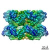 8658MC 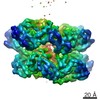 8659C 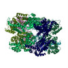 5vcaC M: map data used to model this data C: citing same article ( |
|---|---|
| Similar structure data |
-Links
 PDBj
PDBj


- Assembly
| Deposited unit | 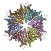
|
|---|---|
| 1 |
|
-Components
| #1: Protein | Mass: 62996.246 Da / Num. of mol.: 6 / Fragment: UNP residues 183-745 Source method: isolated from a genetically manipulated source Source: (gene. exp.) Thermoplasma acidophilum (strain ATCC 25905 / DSM 1728 / JCM 9062 / NBRC 15155 / AMRC-C165) (acidophilic)Strain: ATCC 25905 / DSM 1728 / JCM 9062 / NBRC 15155 / AMRC-C165 Gene: vat, Ta0840 / Plasmid: PPROEX / Production host:  #2: Chemical | ChemComp-ATP /   Mass: 507.181 Da / Num. of mol.: 12 / Source method: obtained synthetically / Formula: C10H16N5O13P3 / Comment: ATP, energy-carrying molecule*YM Mass: 507.181 Da / Num. of mol.: 12 / Source method: obtained synthetically / Formula: C10H16N5O13P3 / Comment: ATP, energy-carrying molecule*YMHas protein modification | Y | |
|---|
-Experimental details
-Experiment
| Experiment | Method: ELECTRON MICROSCOPY |
|---|---|
| EM experiment | Aggregation state: PARTICLE / 3D reconstruction method: single particle reconstruction |
- Sample preparation
| Component | Name: Structural basis for substrate unfolding by the VCP like ATPase from T. Acidophilum Type: COMPLEX Details: Stacked-ring state, ATPgS loaded N-terminal domain removed Entity ID: #1 / Source: RECOMBINANT |
|---|---|
| Molecular weight | Value: 0.38 MDa / Experimental value: NO |
| Source (natural) | Organism: Thermoplasma acidophilum (acidophilic) |
| Source (recombinant) | Organism: |
| Buffer solution | pH: 7.5 |
| Specimen | Conc.: 20 mg/ml / Embedding applied: NO / Shadowing applied: NO / Staining applied: NO / Vitrification applied: YES |
| Specimen support | Grid material: COPPER/RHODIUM / Grid mesh size: 400 divisions/in. / Grid type: Electron Microscopy Sciences M400 |
| Vitrification | Instrument: FEI VITROBOT MARK III / Cryogen name: ETHANE-PROPANE / Humidity: 100 % / Chamber temperature: 277 K / Details: Blot for 4 seconds before plunging |
- Electron microscopy imaging
| Microscopy | Model: FEI TECNAI 20 |
|---|---|
| Electron gun | Electron source:  FIELD EMISSION GUN / Accelerating voltage: 200 kV / Illumination mode: FLOOD BEAM FIELD EMISSION GUN / Accelerating voltage: 200 kV / Illumination mode: FLOOD BEAM |
| Electron lens | Mode: BRIGHT FIELD / Nominal magnification: 25000 X / Nominal defocus max: 2900 nm / Nominal defocus min: 1700 nm / Cs: 2 mm / C2 aperture diameter: 30 µm / Alignment procedure: COMA FREE |
| Specimen holder | Cryogen: NITROGEN Specimen holder model: GATAN 626 SINGLE TILT LIQUID NITROGEN CRYO TRANSFER HOLDER |
| Image recording | Average exposure time: 15 sec. / Electron dose: 35 e/Å2 / Detector mode: COUNTING / Film or detector model: GATAN K2 SUMMIT (4k x 4k) / Num. of grids imaged: 3 / Num. of real images: 246 |
| Image scans | Width: 3838 / Height: 3710 / Movie frames/image: 30 / Used frames/image: 1-30 |
- Processing
| EM software |
| ||||||||||||||||||||||||||||||||||||||||||||||||||
|---|---|---|---|---|---|---|---|---|---|---|---|---|---|---|---|---|---|---|---|---|---|---|---|---|---|---|---|---|---|---|---|---|---|---|---|---|---|---|---|---|---|---|---|---|---|---|---|---|---|---|---|
| CTF correction | Type: PHASE FLIPPING AND AMPLITUDE CORRECTION | ||||||||||||||||||||||||||||||||||||||||||||||||||
| Particle selection | Num. of particles selected: 171381 | ||||||||||||||||||||||||||||||||||||||||||||||||||
| Symmetry | Point symmetry: C6 (6 fold cyclic) | ||||||||||||||||||||||||||||||||||||||||||||||||||
| 3D reconstruction | Resolution: 3.9 Å / Resolution method: FSC 0.143 CUT-OFF / Num. of particles: 75205 / Algorithm: FOURIER SPACE / Num. of class averages: 1 / Symmetry type: POINT | ||||||||||||||||||||||||||||||||||||||||||||||||||
| Atomic model building | Protocol: FLEXIBLE FIT / Space: REAL |
