 Movie
Movie Controller

[English] 日本語
 Yorodumi
- PDB-5v3c: Crystal structure of TGT in complex with 4-(aminomethane)cyclohex... -
+ Open data

- Basic information
| Entry | Database: PDB / ID: 5v3c | ||||||
|---|---|---|---|---|---|---|---|
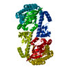
| Title | Crystal structure of TGT in complex with 4-(aminomethane)cyclohexane-1-carboxylic acid | ||||||
 Components Components | Queuine tRNA-ribosyltransferase | ||||||
 Keywords Keywords | TRANSFERASE / TGT / TRNA / Guanine exchange enzyme / transglycosylase | ||||||
| Function / homology |  Function and homology information Function and homology informationtRNA-guanosine34 preQ1 transglycosylase / tRNA-guanosine(34) queuine transglycosylase activity / tRNA queuosine(34) biosynthetic process / metal ion binding / cytosol Similarity search - Function | ||||||
| Biological species |  Zymomonas mobilis (bacteria) Zymomonas mobilis (bacteria) | ||||||
| Method |  X-RAY DIFFRACTION / SYNCHROTRON / MOLECULAR REPLACEMENT / Resolution: 1.419 Å X-RAY DIFFRACTION / SYNCHROTRON / MOLECULAR REPLACEMENT / Resolution: 1.419 Å | ||||||
 Authors Authors | Hassaan, E. / Heine, A. / Klebe, G. | ||||||
 Citation Citation | Journal: Chemmedchem / Year: 2020 Title: Fragments as Novel Starting Points for tRNA-Guanine Transglycosylase Inhibitors Found by Alternative Screening Strategies. Authors: Hassaan, E. / Eriksson, P.O. / Geschwindner, S. / Heine, A. / Klebe, G. | ||||||
| History |
|
- Structure visualization
| Structure viewer | Molecule: MolmilJmol/JSmol |
|---|
- Downloads & links
-Download
| PDBx/mmCIF format | 5v3c.cif.gz | 222.2 KB | Display | PDBx/mmCIF format |
|---|---|---|---|---|
| PDB format | pdb5v3c.ent.gz | 178.5 KB | Display | PDB format |
| PDBx/mmJSON format | 5v3c.json.gz | Tree view | PDBx/mmJSON format | |
| Others |  Other downloads Other downloads |
-Validation report
| Arichive directory | https://data.pdbj.org/pub/pdb/validation_reports/v3/5v3cftp://data.pdbj.org/pub/pdb/validation_reports/v3/5v3c | HTTPS FTP |
|---|
-Related structure data
| Related structure data |  5n6fC  5sw3C  5utiC  5utjC  6fsoC  4lbuS S: Starting model for refinement C: citing same article ( |
|---|---|
| Similar structure data |


-Links
 PDBj
PDBj- Assembly
| Deposited unit | 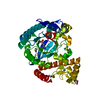
| ||||||||
|---|---|---|---|---|---|---|---|---|---|
| 1 | 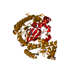
| ||||||||
| Unit cell |
|
-Components
-Protein , 1 types, 1 molecules A
| #1: Protein | Mass: 41763.484 Da / Num. of mol.: 1 Source method: isolated from a genetically manipulated source Source: (gene. exp.) Zymomonas mobilis (bacteria) / Gene: tgt, ZMO0363 / Plasmid: PPR-IBA2 / Production host: References: UniProt: P28720, tRNA-guanosine34 preQ1 transglycosylase |
|---|
-Non-polymers , 6 types, 228 molecules 


| #2: Chemical | ChemComp-ZN /  Mass: 65.409 Da / Num. of mol.: 1 / Source method: obtained synthetically / Formula: Zn Mass: 65.409 Da / Num. of mol.: 1 / Source method: obtained synthetically / Formula: Zn | ||||||
|---|---|---|---|---|---|---|---|
| #3: Chemical | ChemComp-AMH /  Mass: 157.210 Da / Num. of mol.: 1 / Source method: obtained synthetically / Formula: C8H15NO2 Mass: 157.210 Da / Num. of mol.: 1 / Source method: obtained synthetically / Formula: C8H15NO2 | ||||||
| #4: Chemical |  Mass: 106.120 Da / Num. of mol.: 2 / Source method: obtained synthetically / Formula: C4H10O3 Mass: 106.120 Da / Num. of mol.: 2 / Source method: obtained synthetically / Formula: C4H10O3#5: Chemical |  Mass: 78.133 Da / Num. of mol.: 2 / Source method: obtained synthetically / Formula: C2H6OS / Comment: DMSO, precipitant*YM Mass: 78.133 Da / Num. of mol.: 2 / Source method: obtained synthetically / Formula: C2H6OS / Comment: DMSO, precipitant*YM#6: Chemical | ChemComp-PGE / |  Mass: 150.173 Da / Num. of mol.: 1 / Source method: isolated from a natural source / Formula: C6H14O4 Mass: 150.173 Da / Num. of mol.: 1 / Source method: isolated from a natural source / Formula: C6H14O4#7: Water | ChemComp-HOH / | Mass: 18.015 Da / Num. of mol.: 221 / Source method: isolated from a natural source / Formula: H2O |
-Experimental details
-Experiment
| Experiment | Method: X-RAY DIFFRACTION / Number of used crystals: 1 |
|---|
- Sample preparation
| Crystal | Density Matthews: 2.5 Å3/Da / Density % sol: 50.9 % |
|---|---|
| Crystal grow | Temperature: 289 K / Method: vapor diffusion, sitting drop / pH: 5.5 / Details: 13% PEG 8000, 100MM MES, 1MM DTT, 10% DMSO |
-Data collection
| Diffraction | Mean temperature: 100 K |
|---|---|
| Diffraction source | Source: SYNCHROTRON / Site: BESSY  / Beamline: 14.1 / Wavelength: 0.91841 Å / Beamline: 14.1 / Wavelength: 0.91841 Å |
| Detector | Type: DECTRIS PILATUS 6M / Detector: PIXEL / Date: Apr 22, 2016 |
| Radiation | Protocol: SINGLE WAVELENGTH / Monochromatic (M) / Laue (L): M / Scattering type: x-ray |
| Radiation wavelength | Wavelength: 0.91841 Å / Relative weight: 1 |
| Reflection | Resolution: 1.419→44.328 Å / Num. obs: 73715 / % possible obs: 99.4 % / Redundancy: 3.7 % / Rsym value: 0.046 / Net I/σ(I): 15.59 |
| Reflection shell | Resolution: 1.42→1.51 Å / Redundancy: 3.7 % / Rmerge(I) obs: 0.551 / Mean I/σ(I) obs: 2.06 / % possible all: 98.4 |
- Processing
| Software |
| |||||||||||||||||||||||||||||||||||||||||||||||||||||||||||||||||||||||||||||||||||||||||||||||||||||||||||||||||||||||||||||||||||||||||||||||||||||||||||||||||||||||||||||||||||||||||||||
|---|---|---|---|---|---|---|---|---|---|---|---|---|---|---|---|---|---|---|---|---|---|---|---|---|---|---|---|---|---|---|---|---|---|---|---|---|---|---|---|---|---|---|---|---|---|---|---|---|---|---|---|---|---|---|---|---|---|---|---|---|---|---|---|---|---|---|---|---|---|---|---|---|---|---|---|---|---|---|---|---|---|---|---|---|---|---|---|---|---|---|---|---|---|---|---|---|---|---|---|---|---|---|---|---|---|---|---|---|---|---|---|---|---|---|---|---|---|---|---|---|---|---|---|---|---|---|---|---|---|---|---|---|---|---|---|---|---|---|---|---|---|---|---|---|---|---|---|---|---|---|---|---|---|---|---|---|---|---|---|---|---|---|---|---|---|---|---|---|---|---|---|---|---|---|---|---|---|---|---|---|---|---|---|---|---|---|---|---|---|---|
| Refinement | Method to determine structure: MOLECULAR REPLACEMENT Starting model: 4LBU Resolution: 1.419→44.328 Å / SU ML: 0.14 / Cross valid method: FREE R-VALUE / σ(F): 1.35 / Phase error: 14.58 / Stereochemistry target values: ML
| |||||||||||||||||||||||||||||||||||||||||||||||||||||||||||||||||||||||||||||||||||||||||||||||||||||||||||||||||||||||||||||||||||||||||||||||||||||||||||||||||||||||||||||||||||||||||||||
| Solvent computation | Shrinkage radii: 0.9 Å / VDW probe radii: 1.11 Å / Solvent model: FLAT BULK SOLVENT MODEL | |||||||||||||||||||||||||||||||||||||||||||||||||||||||||||||||||||||||||||||||||||||||||||||||||||||||||||||||||||||||||||||||||||||||||||||||||||||||||||||||||||||||||||||||||||||||||||||
| Refinement step | Cycle: LAST / Resolution: 1.419→44.328 Å
| |||||||||||||||||||||||||||||||||||||||||||||||||||||||||||||||||||||||||||||||||||||||||||||||||||||||||||||||||||||||||||||||||||||||||||||||||||||||||||||||||||||||||||||||||||||||||||||
| Refine LS restraints |
| |||||||||||||||||||||||||||||||||||||||||||||||||||||||||||||||||||||||||||||||||||||||||||||||||||||||||||||||||||||||||||||||||||||||||||||||||||||||||||||||||||||||||||||||||||||||||||||
| LS refinement shell |
|
