 Movie
Movie Controller


+ Open data

- Basic information
| Entry | Database: PDB / ID: 5v2h | |||||||||||||||||||||||
|---|---|---|---|---|---|---|---|---|---|---|---|---|---|---|---|---|---|---|---|---|---|---|---|---|
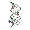
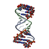
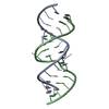
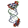
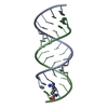
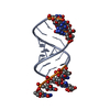
| Title | RNA octamer containing glycol nucleic acid, SgnT | |||||||||||||||||||||||
 Components Components | RNA (5'-R(* Keywords KeywordsRNA / glycol nucleic acid | Function / homology | COBALT HEXAMMINE(III) / RNA |  Function and homology information Function and homology informationBiological species | synthetic construct (others) | Method |  X-RAY DIFFRACTION / SYNCHROTRON / SAD / Resolution: 1.08001306231 Å X-RAY DIFFRACTION / SYNCHROTRON / SAD / Resolution: 1.08001306231 Å  Authors AuthorsHarp, J.M. / Egli, M. |  CitationJournal: J. Am. Chem. Soc. / Year: 2017 CitationJournal: J. Am. Chem. Soc. / Year: 2017Title: Chirality Dependent Potency Enhancement and Structural Impact of Glycol Nucleic Acid Modification on siRNA. Authors: Schlegel, M.K. / Foster, D.J. / Kel'in, A.V. / Zlatev, I. / Bisbe, A. / Jayaraman, M. / Lackey, J.G. / Rajeev, K.G. / Charisse, K. / Harp, J. / Pallan, P.S. / Maier, M.A. / Egli, M. / Manoharan, M. History |
|
- Structure visualization
| Structure viewer | Molecule: MolmilJmol/JSmol |
|---|
- Downloads & links
-Download
| PDBx/mmCIF format | 5v2h.cif.gz | 72.5 KB | Display | PDBx/mmCIF format |
|---|---|---|---|---|
| PDB format | pdb5v2h.ent.gz | 47.8 KB | Display | PDB format |
| PDBx/mmJSON format | 5v2h.json.gz | Tree view | PDBx/mmJSON format | |
| Others |  Other downloads Other downloads |
-Validation report
| Arichive directory | https://data.pdbj.org/pub/pdb/validation_reports/v2/5v2hftp://data.pdbj.org/pub/pdb/validation_reports/v2/5v2h | HTTPS FTP |
|---|
-Related structure data


-Links
 PDBj
PDBj
- Assembly
| Deposited unit | 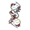
| ||||||||||||
|---|---|---|---|---|---|---|---|---|---|---|---|---|---|
| 1 |
| ||||||||||||
| Unit cell |
| ||||||||||||
| Components on special symmetry positions |
|
-Components
| #1: RNA chain | Mass: 2561.448 Da / Num. of mol.: 4 / Source method: obtained synthetically / Source: (synth.) synthetic construct (others) #2: Chemical | ChemComp-NCO / |   Mass: 161.116 Da / Num. of mol.: 1 / Source method: obtained synthetically / Formula: CoH18N6 Mass: 161.116 Da / Num. of mol.: 1 / Source method: obtained synthetically / Formula: CoH18N6#3: Chemical | ChemComp-MG / |   Mass: 24.305 Da / Num. of mol.: 1 / Source method: obtained synthetically / Formula: Mg Mass: 24.305 Da / Num. of mol.: 1 / Source method: obtained synthetically / Formula: Mg#4: Water | ChemComp-HOH / |  Mass: 18.015 Da / Num. of mol.: 247 / Source method: isolated from a natural source / Formula: H2O Mass: 18.015 Da / Num. of mol.: 247 / Source method: isolated from a natural source / Formula: H2O |
|---|
-Experimental details
-Experiment
| Experiment | Method: X-RAY DIFFRACTION / Number of used crystals: 1 |
|---|
- Sample preparation
| Crystal | Density Matthews: 2.08 Å3/Da / Density % sol: 40.81 % |
|---|---|
| Crystal grow | Temperature: 291 K / Method: vapor diffusion, sitting drop / pH: 5.5 Details: 0.5 mM oligonucleotide, 40 mM sodium chloride, 10 mM magnesium chloride, 10 mM cobalt(III) hexamine chloride, 20 mM sodium cacodylate, 5% 2-methyl-2,4-pentanediol equilibrated against 40% 2- ...Details: 0.5 mM oligonucleotide, 40 mM sodium chloride, 10 mM magnesium chloride, 10 mM cobalt(III) hexamine chloride, 20 mM sodium cacodylate, 5% 2-methyl-2,4-pentanediol equilibrated against 40% 2-methyl-2-4-pentanediol |
-Data collection
| Diffraction | Mean temperature: 100 K |
|---|---|
| Diffraction source | Source: SYNCHROTRON / Site: APS  / Beamline: 21-ID-D / Wavelength: 0.91836 Å / Beamline: 21-ID-D / Wavelength: 0.91836 Å |
| Detector | Type: DECTRIS EIGER X 9M / Detector: PIXEL / Date: Mar 19, 2016 |
| Radiation | Protocol: SINGLE WAVELENGTH / Monochromatic (M) / Laue (L): M / Scattering type: x-ray |
| Radiation wavelength | Wavelength: 0.91836 Å / Relative weight: 1 |
| Reflection | Resolution: 1.08→33.781 Å / Num. obs: 35479 / % possible obs: 97.4 % / Redundancy: 6.3 % / Biso Wilson estimate: 5.18 Å2 / CC1/2: 0.969 / Rmerge(I) obs: 0.164 / Rpim(I) all: 0.069 / Net I/σ(I): 10.74 |
| Reflection shell | Resolution: 1.08→1.119 Å / Redundancy: 2 % / Rmerge(I) obs: 0.2652 / Mean I/σ(I) obs: 2.44 / Num. unique obs: 3532 / CC1/2: 0.822 / Rpim(I) all: 0.2652 / % possible all: 97.14 |
- Processing
| Software |
| ||||||||||||||||||||||||||||||||||||||||||||||||||||||||||||||||||||||||||||||||||||||||||||||||||
|---|---|---|---|---|---|---|---|---|---|---|---|---|---|---|---|---|---|---|---|---|---|---|---|---|---|---|---|---|---|---|---|---|---|---|---|---|---|---|---|---|---|---|---|---|---|---|---|---|---|---|---|---|---|---|---|---|---|---|---|---|---|---|---|---|---|---|---|---|---|---|---|---|---|---|---|---|---|---|---|---|---|---|---|---|---|---|---|---|---|---|---|---|---|---|---|---|---|---|---|
| Refinement | Method to determine structure: SAD / Resolution: 1.08001306231→33.7809065231 Å / SU ML: 0.0713631076248 / Cross valid method: FREE R-VALUE / σ(F): 1.42213021666 / Phase error: 15.8804008545
| ||||||||||||||||||||||||||||||||||||||||||||||||||||||||||||||||||||||||||||||||||||||||||||||||||
| Solvent computation | Shrinkage radii: 0.9 Å / VDW probe radii: 1.11 Å | ||||||||||||||||||||||||||||||||||||||||||||||||||||||||||||||||||||||||||||||||||||||||||||||||||
| Displacement parameters | Biso mean: 11.8546699308 Å2 | ||||||||||||||||||||||||||||||||||||||||||||||||||||||||||||||||||||||||||||||||||||||||||||||||||
| Refinement step | Cycle: LAST / Resolution: 1.08001306231→33.7809065231 Å
| ||||||||||||||||||||||||||||||||||||||||||||||||||||||||||||||||||||||||||||||||||||||||||||||||||
| Refine LS restraints |
| ||||||||||||||||||||||||||||||||||||||||||||||||||||||||||||||||||||||||||||||||||||||||||||||||||
| LS refinement shell |
|
