 Movie
Movie Controller

[English] 日本語
 Yorodumi
- PDB-6owl: RNA oligonucleotides with 3'-arabino guanosine co-crystallized wi... -
+ Open data

- Basic information
| Entry | Database: PDB / ID: 6owl | |||||||||
|---|---|---|---|---|---|---|---|---|---|---|
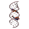
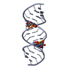
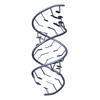
| Title | RNA oligonucleotides with 3'-arabino guanosine co-crystallized with GMP | |||||||||
 Components Components |
| |||||||||
 Keywords Keywords | RNA / arabino | |||||||||
| Function / homology | RNA / RNA (> 10) Function and homology information Function and homology information | |||||||||
| Biological species | synthetic construct (others) | |||||||||
| Method |  X-RAY DIFFRACTION / SYNCHROTRON / MOLECULAR REPLACEMENT / Resolution: 2 Å X-RAY DIFFRACTION / SYNCHROTRON / MOLECULAR REPLACEMENT / Resolution: 2 Å | |||||||||
 Authors Authors | Szostak, J.W. / Kim, S. / Zhang, W. | |||||||||
| Funding support |  United States, 1items United States, 1items
| |||||||||
 Citation Citation | Journal: J.Am.Chem.Soc. / Year: 2020 Title: A Model for the Emergence of RNA from a Prebiotically Plausible Mixture of Ribonucleotides, Arabinonucleotides, and 2'-Deoxynucleotides. Authors: Kim, S.C. / Zhou, L. / Zhang, W. / O'Flaherty, D.K. / Rondo-Brovetto, V. / Szostak, J.W. | |||||||||
| History |
|
- Structure visualization
| Structure viewer | Molecule: MolmilJmol/JSmol |
|---|
- Downloads & links
-Download
| PDBx/mmCIF format | 6owl.cif.gz | 22.1 KB | Display | PDBx/mmCIF format |
|---|---|---|---|---|
| PDB format | pdb6owl.ent.gz | 13.2 KB | Display | PDB format |
| PDBx/mmJSON format | 6owl.json.gz | Tree view | PDBx/mmJSON format | |
| Others |  Other downloads Other downloads |
-Validation report
| Arichive directory | https://data.pdbj.org/pub/pdb/validation_reports/ow/6owlftp://data.pdbj.org/pub/pdb/validation_reports/ow/6owl | HTTPS FTP |
|---|
-Related structure data
| Related structure data | 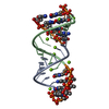 6c8dS S: Starting model for refinement |
|---|---|
| Similar structure data |

-Links
 PDBj
PDBj


- Assembly
| Deposited unit | 
| ||||||||
|---|---|---|---|---|---|---|---|---|---|
| 1 | 
| ||||||||
| Unit cell |
|
-Components
| #1: RNA chain | Mass: 4512.804 Da / Num. of mol.: 1 Source method: isolated from a genetically manipulated source Source: (gene. exp.) synthetic construct (others) / Production host: synthetic construct (others) | ||||
|---|---|---|---|---|---|
| #2: RNA chain | Mass: 645.454 Da / Num. of mol.: 2 Source method: isolated from a genetically manipulated source Source: (gene. exp.) synthetic construct (others) / Production host: synthetic construct (others) #3: Water | ChemComp-HOH / |  Mass: 18.015 Da / Num. of mol.: 8 / Source method: isolated from a natural source / Formula: H2O Mass: 18.015 Da / Num. of mol.: 8 / Source method: isolated from a natural source / Formula: H2OHas protein modification | N | |
-Experimental details
-Experiment
| Experiment | Method: X-RAY DIFFRACTION / Number of used crystals: 1 |
|---|
- Sample preparation
| Crystal | Density Matthews: 2 Å3/Da / Density % sol: 38.47 % |
|---|---|
| Crystal grow | Temperature: 293 K / Method: vapor diffusion, sitting drop / pH: 6 Details: 0.08 M Sodium chloride, 0.012 M Potassium chloride, 0.02 M Magnesium chloride hexahydrate, 0.04 M Sodium cacodylate trihydrate pH 6.0, 30% v/v (+/-)-2-Methyl-2,4-pentanediol, 0.012 M Spermine tetrahydrochloride |
-Data collection
| Diffraction | Mean temperature: 100 K / Serial crystal experiment: N |
|---|---|
| Diffraction source | Source: SYNCHROTRON / Site: ALS / Beamline: 8.2.2 / Wavelength: 1 Å |
| Detector | Type: MAR CCD 130 mm / Detector: CCD / Date: Mar 29, 2019 |
| Radiation | Protocol: SINGLE WAVELENGTH / Monochromatic (M) / Laue (L): M / Scattering type: x-ray |
| Radiation wavelength | Wavelength: 1 Å / Relative weight: 1 |
| Reflection | Resolution: 2→50 Å / Num. obs: 3409 / % possible obs: 99.7 % / Redundancy: 7.1 % / CC1/2: 0.982 / Rmerge(I) obs: 0.082 / Rrim(I) all: 0.09 / Χ2: 0.831 / Net I/σ(I): 19.3 |
| Reflection shell | Resolution: 2→2.07 Å / Redundancy: 5.3 % / Rmerge(I) obs: 0.564 / Mean I/σ(I) obs: 2.3 / Num. unique obs: 332 / CC1/2: 0.94 / Rrim(I) all: 0.627 / Χ2: 0.539 / % possible all: 100 |
- Processing
| Software |
| ||||||||||||||||||||||||||||||||||||||||||||||||||||||||||||||||||||||||||||||||||||||||||||||||||||||||||||||||||||||||||||||||||||||||||||||||||||||||||||||||||||||||||||||||||||||
|---|---|---|---|---|---|---|---|---|---|---|---|---|---|---|---|---|---|---|---|---|---|---|---|---|---|---|---|---|---|---|---|---|---|---|---|---|---|---|---|---|---|---|---|---|---|---|---|---|---|---|---|---|---|---|---|---|---|---|---|---|---|---|---|---|---|---|---|---|---|---|---|---|---|---|---|---|---|---|---|---|---|---|---|---|---|---|---|---|---|---|---|---|---|---|---|---|---|---|---|---|---|---|---|---|---|---|---|---|---|---|---|---|---|---|---|---|---|---|---|---|---|---|---|---|---|---|---|---|---|---|---|---|---|---|---|---|---|---|---|---|---|---|---|---|---|---|---|---|---|---|---|---|---|---|---|---|---|---|---|---|---|---|---|---|---|---|---|---|---|---|---|---|---|---|---|---|---|---|---|---|---|---|---|
| Refinement | Method to determine structure: MOLECULAR REPLACEMENT Starting model: 6C8D Resolution: 2→22 Å / Cor.coef. Fo:Fc: 0.955 / Cor.coef. Fo:Fc free: 0.894 / SU B: 6.49 / SU ML: 0.188 / Cross valid method: THROUGHOUT / ESU R: 0.213 / ESU R Free: 0.213 / Details: HYDROGENS HAVE BEEN ADDED IN THE RIDING POSITIONS
| ||||||||||||||||||||||||||||||||||||||||||||||||||||||||||||||||||||||||||||||||||||||||||||||||||||||||||||||||||||||||||||||||||||||||||||||||||||||||||||||||||||||||||||||||||||||
| Solvent computation | Ion probe radii: 0.8 Å / Shrinkage radii: 0.8 Å / VDW probe radii: 1.2 Å | ||||||||||||||||||||||||||||||||||||||||||||||||||||||||||||||||||||||||||||||||||||||||||||||||||||||||||||||||||||||||||||||||||||||||||||||||||||||||||||||||||||||||||||||||||||||
| Displacement parameters | Biso mean: 41.933 Å2
| ||||||||||||||||||||||||||||||||||||||||||||||||||||||||||||||||||||||||||||||||||||||||||||||||||||||||||||||||||||||||||||||||||||||||||||||||||||||||||||||||||||||||||||||||||||||
| Refinement step | Cycle: 1 / Resolution: 2→22 Å
| ||||||||||||||||||||||||||||||||||||||||||||||||||||||||||||||||||||||||||||||||||||||||||||||||||||||||||||||||||||||||||||||||||||||||||||||||||||||||||||||||||||||||||||||||||||||
| Refine LS restraints |
|