 Movie
Movie Controller

[English] 日本語
 Yorodumi
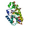
- PDB-5u9j: Crystal structure of the FKBP domain of human aryl hydrocarbon re... -
+ Open data

- Basic information
| Entry | Database: PDB / ID: 5u9j | |||||||||
|---|---|---|---|---|---|---|---|---|---|---|
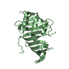
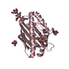
| Title | Crystal structure of the FKBP domain of human aryl hydrocarbon receptor-interacting protein-like 1 (AIPL1) complexed with geranyl geranyl pyrophoshate | |||||||||
 Components Components | Aryl hydrocarbon receptor-interacting protein-like 1 (AIPL1) | |||||||||
 Keywords Keywords | SIGNALING PROTEIN / AIPL1 / FKBP / chaperone / PDE6 / photoreceptor / LCA / isoprenyl | |||||||||
| Function / homology |  Function and homology information Function and homology informationfarnesylated protein binding / protein farnesylation / regulation of opsin-mediated signaling pathway / retina homeostasis / phototransduction, visible light / photoreceptor inner segment / visual perception / peptidyl-prolyl cis-trans isomerase activity / : / nuclear speck ...farnesylated protein binding / protein farnesylation / regulation of opsin-mediated signaling pathway / retina homeostasis / phototransduction, visible light / photoreceptor inner segment / visual perception / peptidyl-prolyl cis-trans isomerase activity / : / nuclear speck / apoptotic process / negative regulation of apoptotic process / nucleoplasm / nucleus / cytoplasm / cytosol Similarity search - Function | |||||||||
| Biological species |  Homo sapiens (human) Homo sapiens (human) | |||||||||
| Method |  X-RAY DIFFRACTION / SYNCHROTRON / MOLECULAR REPLACEMENT / Resolution: 2.1 Å X-RAY DIFFRACTION / SYNCHROTRON / MOLECULAR REPLACEMENT / Resolution: 2.1 Å | |||||||||
 Authors Authors | Yadav, R.P. / Gakhar, L. / Liping, Y. / Artemyev, N.O. | |||||||||
| Funding support |  United States, 1items United States, 1items
| |||||||||
 Citation Citation | Journal: Proc. Natl. Acad. Sci. U.S.A. / Year: 2017 Title: Unique structural features of the AIPL1-FKBP domain that support prenyl lipid binding and underlie protein malfunction in blindness. Authors: Yadav, R.P. / Gakhar, L. / Yu, L. / Artemyev, N.O. | |||||||||
| History |
|
- Structure visualization
| Structure viewer | Molecule: MolmilJmol/JSmol |
|---|
- Downloads & links
-Download
| PDBx/mmCIF format | 5u9j.cif.gz | 131.5 KB | Display | PDBx/mmCIF format |
|---|---|---|---|---|
| PDB format | pdb5u9j.ent.gz | 103.5 KB | Display | PDB format |
| PDBx/mmJSON format | 5u9j.json.gz | Tree view | PDBx/mmJSON format | |
| Others |  Other downloads Other downloads |
-Validation report
| Arichive directory | https://data.pdbj.org/pub/pdb/validation_reports/u9/5u9jftp://data.pdbj.org/pub/pdb/validation_reports/u9/5u9j | HTTPS FTP |
|---|
-Related structure data
| Related structure data | 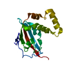 5u9aSC 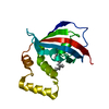 5u9iC 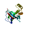 5u9kC  5v35C S: Starting model for refinement C: citing same article ( |
|---|---|
| Similar structure data |


-Links
 PDBj
PDBj


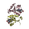
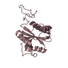
- Assembly
| Deposited unit | 
| ||||||||
|---|---|---|---|---|---|---|---|---|---|
| 1 | 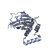
| ||||||||
| 2 | 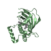
| ||||||||
| Unit cell |
| ||||||||
| Components on special symmetry positions |
|
-Components
| #1: Protein | Mass: 19327.109 Da / Num. of mol.: 2 / Fragment: UNP residues 2-161 Source method: isolated from a genetically manipulated source Source: (gene. exp.) Homo sapiens (human) / Gene: AIPL1, AIPL2 / Plasmid: pET-15b / Production host:  #2: Chemical | 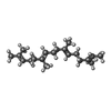  Mass: 274.484 Da / Num. of mol.: 2 / Source method: isolated from a natural source / Formula: C20H34 Mass: 274.484 Da / Num. of mol.: 2 / Source method: isolated from a natural source / Formula: C20H34#3: Chemical | ChemComp-IPA / |   Mass: 60.095 Da / Num. of mol.: 1 / Source method: obtained synthetically / Formula: C3H8O Mass: 60.095 Da / Num. of mol.: 1 / Source method: obtained synthetically / Formula: C3H8O#4: Chemical | ChemComp-NA / |   Mass: 22.990 Da / Num. of mol.: 1 / Source method: obtained synthetically / Formula: Na Mass: 22.990 Da / Num. of mol.: 1 / Source method: obtained synthetically / Formula: Na#5: Water | ChemComp-HOH / |  Mass: 18.015 Da / Num. of mol.: 141 / Source method: isolated from a natural source / Formula: H2O Mass: 18.015 Da / Num. of mol.: 141 / Source method: isolated from a natural source / Formula: H2O |
|---|
-Experimental details
-Experiment
| Experiment | Method: X-RAY DIFFRACTION / Number of used crystals: 1 |
|---|
- Sample preparation
| Crystal | Density Matthews: 2.45 Å3/Da / Density % sol: 49.82 % |
|---|---|
| Crystal grow | Temperature: 291 K / Method: vapor diffusion, hanging drop Details: 100 mM Na-Citrate 20%(W/V) PEG 4000 20% (V/V) 2-Propanol PH range: 5-7 |
-Data collection
| Diffraction | Mean temperature: 100 K |
|---|---|
| Diffraction source | Source: SYNCHROTRON / Site: ALS / Beamline: 4.2.2 / Wavelength: 1 Å |
| Detector | Type: RDI CMOS_8M / Detector: CMOS / Date: Oct 11, 2016 |
| Radiation | Protocol: SINGLE WAVELENGTH / Monochromatic (M) / Laue (L): M / Scattering type: x-ray |
| Radiation wavelength | Wavelength: 1 Å / Relative weight: 1 |
| Reflection | Resolution: 2.1→41.152 Å / Num. obs: 17642 / % possible obs: 98.1 % / Redundancy: 7 % / CC1/2: 0.999 / Rmerge(I) obs: 0.067 / Net I/σ(I): 20.4 |
| Reflection shell | Resolution: 2.1→2.37 Å / Redundancy: 6.6 % / Rmerge(I) obs: 0.431 / Mean I/σ(I) obs: 5.3 / CC1/2: 0.957 / % possible all: 95.5 |
- Processing
| Software |
| ||||||||||||||||||||||||||||||||||||||||||||||||||||||||||||||||||||||||||||||||||||||||||||||||||
|---|---|---|---|---|---|---|---|---|---|---|---|---|---|---|---|---|---|---|---|---|---|---|---|---|---|---|---|---|---|---|---|---|---|---|---|---|---|---|---|---|---|---|---|---|---|---|---|---|---|---|---|---|---|---|---|---|---|---|---|---|---|---|---|---|---|---|---|---|---|---|---|---|---|---|---|---|---|---|---|---|---|---|---|---|---|---|---|---|---|---|---|---|---|---|---|---|---|---|---|
| Refinement | Method to determine structure: MOLECULAR REPLACEMENT Starting model: 5U9A Resolution: 2.1→41.152 Å / SU ML: 0.35 / Cross valid method: FREE R-VALUE / σ(F): 0.82 / Phase error: 32.62 / Stereochemistry target values: ML
| ||||||||||||||||||||||||||||||||||||||||||||||||||||||||||||||||||||||||||||||||||||||||||||||||||
| Solvent computation | Shrinkage radii: 0.9 Å / VDW probe radii: 1.11 Å / Solvent model: FLAT BULK SOLVENT MODEL | ||||||||||||||||||||||||||||||||||||||||||||||||||||||||||||||||||||||||||||||||||||||||||||||||||
| Refinement step | Cycle: LAST / Resolution: 2.1→41.152 Å
| ||||||||||||||||||||||||||||||||||||||||||||||||||||||||||||||||||||||||||||||||||||||||||||||||||
| Refine LS restraints |
| ||||||||||||||||||||||||||||||||||||||||||||||||||||||||||||||||||||||||||||||||||||||||||||||||||
| LS refinement shell |
|
