negative regulation of tyrosine phosphorylation of STAT protein / positive regulation of interleukin-4-mediated signaling pathway / NAD+ catabolic process / Maturation of nucleoprotein / Nicotinate metabolism / positive regulation of tyrosine phosphorylation of STAT protein / Maturation of nucleoprotein / protein poly-ADP-ribosylation / NAD+-protein-glutamate ADP-ribosyltransferase activity / negative regulation of type II interferon-mediated signaling pathway ...negative regulation of tyrosine phosphorylation of STAT protein / positive regulation of interleukin-4-mediated signaling pathway / NAD+ catabolic process / Maturation of nucleoprotein / Nicotinate metabolism / positive regulation of tyrosine phosphorylation of STAT protein / Maturation of nucleoprotein / protein poly-ADP-ribosylation / NAD+-protein-glutamate ADP-ribosyltransferase activity / negative regulation of type II interferon-mediated signaling pathway / NAD+-protein mono-ADP-ribosyltransferase activity / Transferases; Glycosyltransferases; Pentosyltransferases / NAD+ poly-ADP-ribosyltransferase activity / NAD+ binding / nucleotidyltransferase activity / transcription corepressor activity / negative regulation of gene expression / innate immune response / enzyme binding / membrane / nucleus / plasma membrane / cytoplasm / cytosol Similarity search - Function
PARP-14, RNA recognition motif 2 / : / : / : / : / : / : / : / : / Parp14 WWE domain ...PARP-14, RNA recognition motif 2 / : / : / : / : / : / : / : / : / Parp14 WWE domain / PARP14, first type I KH domain / PARP14, second RRM domain / PARP14, second type I KH domain / PARP14, third type I KH domain / PARP14, fourth type I KH domain / PARP14, fifth type I KH domain / PARP14, sixth type I KH domain / : / PAR14-like, first RRM domain / : / PARP14-like, eighth type I KH domain / PARP14, third RRM domain / : / WWE domain superfamily / WWE domain / WWE domain profile. / Phosphoenolpyruvate Carboxykinase; domain 3 - #10 / Phosphoenolpyruvate Carboxykinase; domain 3 / Poly(ADP-ribose) polymerase catalytic domain / Poly(ADP-ribose) polymerase, catalytic domain / PARP catalytic domain profile. / Appr-1"-p processing enzyme / Macro domain / Macro domain profile. / Macro domain / Macro domain-like / Nucleotide-binding alpha-beta plait domain superfamily / Alpha-Beta Complex / Alpha Beta Similarity search - Domain/homology
In the structure databanks used in Yorodumi, some data are registered as the other names, "COVID-19 virus" and "2019-nCoV". Here are the details of the virus and the list of structure data.
Jan 31, 2019. EMDB accession codes are about to change! (news from PDBe EMDB page)
EMDB accession codes are about to change! (news from PDBe EMDB page)
The allocation of 4 digits for EMDB accession codes will soon come to an end. Whilst these codes will remain in use, new EMDB accession codes will include an additional digit and will expand incrementally as the available range of codes is exhausted. The current 4-digit format prefixed with “EMD-” (i.e. EMD-XXXX) will advance to a 5-digit format (i.e. EMD-XXXXX), and so on. It is currently estimated that the 4-digit codes will be depleted around Spring 2019, at which point the 5-digit format will come into force.
The EM Navigator/Yorodumi systems omit the EMD- prefix.
Related info.:Q: What is EMD? / ID/Accession-code notation in Yorodumi/EM Navigator
Yorodumi is a browser for structure data from EMDB, PDB, SASBDB, etc.
This page is also the successor to EM Navigator detail page, and also detail information page/front-end page for Omokage search.
The word "yorodu" (or yorozu) is an old Japanese word meaning "ten thousand". "mi" (miru) is to see.
Related info.:EMDB / PDB / SASBDB / Comparison of 3 databanks / Yorodumi Search / Aug 31, 2016. New EM Navigator & Yorodumi / Yorodumi Papers / Jmol/JSmol / Function and homology information / Changes in new EM Navigator and Yorodumi
 Movie
Movie Controller

 Yorodumi
 Open data
 Basic information
 Components
 Keywords
 Function and homology information
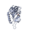
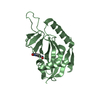
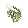
 Homo sapiens (human)
 X-RAY DIFFRACTION /
  Authors
 Citation
 Structure visualization
 Downloads & links
 Other downloads


 PDBj
PDBj Assembly


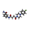
 Mass: 398.431 Da / Num. of mol.: 2 / Source method: obtained synthetically / Formula: C21H23FN4O3
 Mass: 18.015 Da / Num. of mol.: 17 / Source method: isolated from a natural source / Formula: H2O
 Sample preparation
 / Beamline: I03 / Wavelength: 0.97625 Å
 Processing