 Movie
Movie Controller


+ Open data

- Basic information
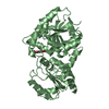
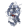
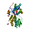
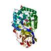
| Entry | Database: PDB / ID: 5u1g | ||||||
|---|---|---|---|---|---|---|---|
| Title | Structure of TP228 ParA-AMPPNP-ParB complex | ||||||
 Components Components |
| ||||||
 Keywords Keywords | CELL CYCLE / REPLICATION / DNA segregation / Walker-box / partition | ||||||
| Function / homology |  Function and homology information Function and homology informationDNA-binding transcription repressor activity / core promoter sequence-specific DNA binding / transcription repressor complex / protein-DNA complex / nucleic acid binding / negative regulation of DNA-templated transcription / identical protein binding Similarity search - Function | ||||||
| Biological species | unidentified plasmid (others) | ||||||
| Method |  X-RAY DIFFRACTION / SYNCHROTRON / MOLECULAR REPLACEMENT / molecular replacement / Resolution: 3.64 Å X-RAY DIFFRACTION / SYNCHROTRON / MOLECULAR REPLACEMENT / molecular replacement / Resolution: 3.64 Å | ||||||
 Authors Authors | Schumacher, M.A. | ||||||
 Citation Citation | Journal: Genes Dev. / Year: 2017 Title: Structures of partition protein ParA with nonspecific DNA and ParB effector reveal molecular insights into principles governing Walker-box DNA segregation. Authors: Zhang, H. / Schumacher, M.A. | ||||||
| History |
|
- Structure visualization
| Structure viewer | Molecule: MolmilJmol/JSmol |
|---|
- Downloads & links
-Download
| PDBx/mmCIF format | 5u1g.cif.gz | 176.3 KB | Display | PDBx/mmCIF format |
|---|---|---|---|---|
| PDB format | pdb5u1g.ent.gz | 140.2 KB | Display | PDB format |
| PDBx/mmJSON format | 5u1g.json.gz | Tree view | PDBx/mmJSON format | |
| Others |  Other downloads Other downloads |
-Validation report
| Arichive directory | https://data.pdbj.org/pub/pdb/validation_reports/u1/5u1gftp://data.pdbj.org/pub/pdb/validation_reports/u1/5u1g | HTTPS FTP |
|---|
-Related structure data
| Related structure data | 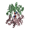 5u1jC 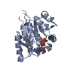 4e07S S: Starting model for refinement C: citing same article ( |
|---|---|
| Similar structure data |


-Links
 PDBj
PDBj


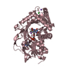
- Assembly
| Deposited unit | 
| ||||||||
|---|---|---|---|---|---|---|---|---|---|
| 1 | 
| ||||||||
| 2 | 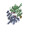
| ||||||||
| Unit cell |
|
-Components
| #1: Protein | Mass: 23062.365 Da / Num. of mol.: 4 Source method: isolated from a genetically manipulated source Source: (gene. exp.) unidentified plasmid (others) / Production host:  #2: Protein/peptide | Mass: 2253.580 Da / Num. of mol.: 4 Source method: isolated from a genetically manipulated source Source: (gene. exp.) unidentified plasmid (others) / Production host: #3: Chemical | ChemComp-ANP /   Mass: 506.196 Da / Num. of mol.: 4 / Source method: obtained synthetically / Formula: C10H17N6O12P3 / Comment: AMP-PNP, energy-carrying molecule analogue*YM Mass: 506.196 Da / Num. of mol.: 4 / Source method: obtained synthetically / Formula: C10H17N6O12P3 / Comment: AMP-PNP, energy-carrying molecule analogue*YM |
|---|
-Experimental details
-Experiment
| Experiment | Method: X-RAY DIFFRACTION / Number of used crystals: 1 |
|---|
- Sample preparation
| Crystal | Density Matthews: 2.7 Å3/Da / Density % sol: 54.42 % |
|---|---|
| Crystal grow | Temperature: 298 K / Method: vapor diffusion, hanging drop Details: 25% PEG 20000, 0.1 M MgCl2, 0.1 M Tris pH 8.5. took 6 months to grow, Mass spec revealed TP228 FL ParB protein had degraded and only N-term region revealed in crystals |
-Data collection
| Diffraction | Mean temperature: 100 K |
|---|---|
| Diffraction source | Source: SYNCHROTRON / Site: ALS  / Beamline: 8.3.1 / Wavelength: 1 Å / Beamline: 8.3.1 / Wavelength: 1 Å |
| Detector | Type: ADSC QUANTUM 210r / Detector: CCD / Date: May 25, 2015 |
| Radiation | Protocol: SINGLE WAVELENGTH / Monochromatic (M) / Laue (L): M / Scattering type: x-ray |
| Radiation wavelength | Wavelength: 1 Å / Relative weight: 1 |
| Reflection | Resolution: 3.65→87.36 Å / Biso Wilson estimate: 59.83 Å2 / CC1/2: 0.98 / Rpim(I) all: 0.127 / Rsym value: 14.5 |
| Reflection shell | Resolution: 3.64→3.85 Å / Redundancy: 2.2 % / Rmerge(I) obs: 0.338 / Mean I/σ(I) obs: 1.9 / CC1/2: 0.843 / % possible all: 78.6 |
-Phasing
| Phasing | Method: molecular replacement |
|---|
- Processing
| Software |
| ||||||||||||||||||||||||||||||||||||||||||||||||||||||||
|---|---|---|---|---|---|---|---|---|---|---|---|---|---|---|---|---|---|---|---|---|---|---|---|---|---|---|---|---|---|---|---|---|---|---|---|---|---|---|---|---|---|---|---|---|---|---|---|---|---|---|---|---|---|---|---|---|---|
| Refinement | Method to determine structure: MOLECULAR REPLACEMENT / Starting model: 40000000 / Resolution: 3.64→73.268 Å / FOM work R set: 0.7793 / SU ML: 0.45 / Cross valid method: FREE R-VALUE / σ(F): 1.39 / Phase error: 27.83 / Stereochemistry target values: ML
| ||||||||||||||||||||||||||||||||||||||||||||||||||||||||
| Solvent computation | Shrinkage radii: 0.9 Å / VDW probe radii: 1.11 Å / Solvent model: FLAT BULK SOLVENT MODEL / Bsol: 20 Å2 / ksol: 0.324 e/Å3 | ||||||||||||||||||||||||||||||||||||||||||||||||||||||||
| Displacement parameters | Biso max: 115.8 Å2 / Biso mean: 56.84 Å2 / Biso min: 6.55 Å2
| ||||||||||||||||||||||||||||||||||||||||||||||||||||||||
| Refinement step | Cycle: final / Resolution: 3.64→73.268 Å
| ||||||||||||||||||||||||||||||||||||||||||||||||||||||||
| Refine LS restraints |
| ||||||||||||||||||||||||||||||||||||||||||||||||||||||||
| LS refinement shell | Refine-ID: X-RAY DIFFRACTION / Rfactor Rfree error: 0 / Total num. of bins used: 7
|
