 Movie
Movie Controller


+ Open data

- Basic information
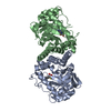
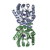
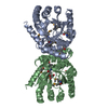
| Entry | Database: PDB / ID: 5u0w | ||||||
|---|---|---|---|---|---|---|---|
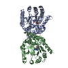
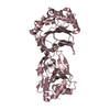
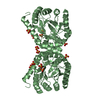
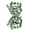
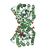
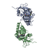
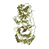
| Title | E. coli dihydropteroate synthase complexed with 9-methylguanine | ||||||
 Components Components | Dihydropteroate synthase | ||||||
 Keywords Keywords | TRANSFERASE/TRANSFERASE INHIBITOR / E. coli / DHPS / complex / pterin site / TRANSFERASE-TRANSFERASE INHIBITOR complex | ||||||
| Function / homology |  Function and homology information Function and homology informationdihydropteroate synthase / dihydropteroate synthase activity / folic acid biosynthetic process / tetrahydrofolate biosynthetic process / metal ion binding / cytosol Similarity search - Function | ||||||
| Biological species |  | ||||||
| Method |  X-RAY DIFFRACTION / SYNCHROTRON / MOLECULAR REPLACEMENT / Resolution: 1.968 Å X-RAY DIFFRACTION / SYNCHROTRON / MOLECULAR REPLACEMENT / Resolution: 1.968 Å | ||||||
 Authors Authors | Chhabra, S. / Dennis, M.L. / Peat, T.S. / Swarbrick, J.D. | ||||||
 Citation Citation | Journal: Chemistry / Year: 2018 Title: 8-Mercaptoguanine Derivatives as Inhibitors of Dihydropteroate Synthase. Authors: Dennis, M.L. / Lee, M.D. / Harjani, J.R. / Ahmed, M. / DeBono, A.J. / Pitcher, N.P. / Wang, Z.C. / Chhabra, S. / Barlow, N. / Rahmani, R. / Cleary, B. / Dolezal, O. / Hattarki, M. / Aurelio, ...Authors: Dennis, M.L. / Lee, M.D. / Harjani, J.R. / Ahmed, M. / DeBono, A.J. / Pitcher, N.P. / Wang, Z.C. / Chhabra, S. / Barlow, N. / Rahmani, R. / Cleary, B. / Dolezal, O. / Hattarki, M. / Aurelio, L. / Shonberg, J. / Graham, B. / Peat, T.S. / Baell, J.B. / Swarbrick, J.D. | ||||||
| History |
|
- Structure visualization
| Structure viewer | Molecule: MolmilJmol/JSmol |
|---|
- Downloads & links
-Download
| PDBx/mmCIF format | 5u0w.cif.gz | 119.8 KB | Display | PDBx/mmCIF format |
|---|---|---|---|---|
| PDB format | pdb5u0w.ent.gz | 91.8 KB | Display | PDB format |
| PDBx/mmJSON format | 5u0w.json.gz | Tree view | PDBx/mmJSON format | |
| Others |  Other downloads Other downloads |
-Validation report
| Arichive directory | https://data.pdbj.org/pub/pdb/validation_reports/u0/5u0wftp://data.pdbj.org/pub/pdb/validation_reports/u0/5u0w | HTTPS FTP |
|---|
-Related structure data
| Related structure data |  5u0vC  5u0yC  5u0zC  5u10C  5u11C  5u12C 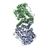 5u13C 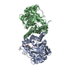 5u14C 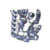 5v79C 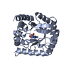 5v7aC 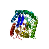 1aj2S  5u0x C: citing same article ( S: Starting model for refinement |
|---|---|
| Similar structure data |


-Links
 PDBj
PDBj

- Assembly
| Deposited unit | 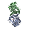
| ||||||||
|---|---|---|---|---|---|---|---|---|---|
| 1 |
| ||||||||
| Unit cell |
|
-Components
| #1: Protein | Mass: 30796.230 Da / Num. of mol.: 2 Source method: isolated from a genetically manipulated source Source: (gene. exp.) Strain: CFT073 / ATCC 700928 / UPEC / Gene: folP, c3933 / Production host: #2: Chemical |   Mass: 165.153 Da / Num. of mol.: 2 / Source method: obtained synthetically / Formula: C6H7N5O Mass: 165.153 Da / Num. of mol.: 2 / Source method: obtained synthetically / Formula: C6H7N5O#3: Chemical | ChemComp-ACY / |   Mass: 60.052 Da / Num. of mol.: 1 / Source method: obtained synthetically / Formula: C2H4O2 Mass: 60.052 Da / Num. of mol.: 1 / Source method: obtained synthetically / Formula: C2H4O2#4: Water | ChemComp-HOH / |  Mass: 18.015 Da / Num. of mol.: 241 / Source method: isolated from a natural source / Formula: H2O Mass: 18.015 Da / Num. of mol.: 241 / Source method: isolated from a natural source / Formula: H2O |
|---|
-Experimental details
-Experiment
| Experiment | Method: X-RAY DIFFRACTION / Number of used crystals: 1 |
|---|
- Sample preparation
| Crystal | Density Matthews: 2.6 Å3/Da / Density % sol: 52.69 % |
|---|---|
| Crystal grow | Temperature: 281 K / Method: vapor diffusion, sitting drop / pH: 6 Details: 16.9% PEG8000 0.1 M sodium cacodylate, pH 6.0 0.143M MgNO3 Protein at 15 mg.mL-1 |
-Data collection
| Diffraction | Mean temperature: 100 K |
|---|---|
| Diffraction source | Source: SYNCHROTRON / Site: Australian Synchrotron  / Beamline: MX1 / Wavelength: 0.9537 Å / Beamline: MX1 / Wavelength: 0.9537 Å |
| Detector | Type: ADSC QUANTUM 210 / Detector: CCD / Date: Oct 20, 2010 |
| Radiation | Protocol: SINGLE WAVELENGTH / Monochromatic (M) / Laue (L): M / Scattering type: x-ray |
| Radiation wavelength | Wavelength: 0.9537 Å / Relative weight: 1 |
| Reflection | Resolution: 1.968→19.771 Å / Num. obs: 44542 / % possible obs: 99.5 % / Redundancy: 7.4 % / Rmerge(I) obs: 0.084 / Net I/σ(I): 13.8 |
- Processing
| Software |
| |||||||||||||||||||||||||||||||||||||||||||||||||||||||||||||||||||||||||||||||||||||||||||||||||||||||||||||||||||||||
|---|---|---|---|---|---|---|---|---|---|---|---|---|---|---|---|---|---|---|---|---|---|---|---|---|---|---|---|---|---|---|---|---|---|---|---|---|---|---|---|---|---|---|---|---|---|---|---|---|---|---|---|---|---|---|---|---|---|---|---|---|---|---|---|---|---|---|---|---|---|---|---|---|---|---|---|---|---|---|---|---|---|---|---|---|---|---|---|---|---|---|---|---|---|---|---|---|---|---|---|---|---|---|---|---|---|---|---|---|---|---|---|---|---|---|---|---|---|---|---|---|
| Refinement | Method to determine structure: MOLECULAR REPLACEMENT Starting model: 1AJ2 Resolution: 1.968→19.771 Å / SU ML: 0.24 / Cross valid method: FREE R-VALUE / σ(F): 1.35 / Phase error: 25.06 / Stereochemistry target values: ML
| |||||||||||||||||||||||||||||||||||||||||||||||||||||||||||||||||||||||||||||||||||||||||||||||||||||||||||||||||||||||
| Solvent computation | Shrinkage radii: 0.9 Å / VDW probe radii: 1.11 Å / Solvent model: FLAT BULK SOLVENT MODEL | |||||||||||||||||||||||||||||||||||||||||||||||||||||||||||||||||||||||||||||||||||||||||||||||||||||||||||||||||||||||
| Refinement step | Cycle: LAST / Resolution: 1.968→19.771 Å
| |||||||||||||||||||||||||||||||||||||||||||||||||||||||||||||||||||||||||||||||||||||||||||||||||||||||||||||||||||||||
| Refine LS restraints |
| |||||||||||||||||||||||||||||||||||||||||||||||||||||||||||||||||||||||||||||||||||||||||||||||||||||||||||||||||||||||
| LS refinement shell |
|
