 Movie
Movie Controller

[English] 日本語
 Yorodumi
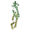
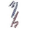
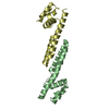
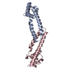
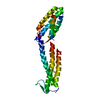
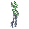
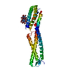
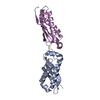
- PDB-5tz0: Structure of the Fremyella diplosiphon Fluorescence Recovery Protein -
+ Open data

- Basic information
| Entry | Database: PDB / ID: 5tz0 | ||||||
|---|---|---|---|---|---|---|---|
| Title | Structure of the Fremyella diplosiphon Fluorescence Recovery Protein | ||||||
 Components Components | Fluorescence Recovery Protein | ||||||
 Keywords Keywords | PROTEIN BINDING / PHOTOPROTECTION / NON-PHOTOCHEMICAL QUENCHING | ||||||
| Function / homology | Fluorescence recovery protein / : / Photoprotection regulator fluorescence recovery protein / thylakoid membrane / Fluorescence recovery protein Function and homology information Function and homology information | ||||||
| Biological species |  Tolypothrix sp. PCC 7601 (bacteria) Tolypothrix sp. PCC 7601 (bacteria) | ||||||
| Method |  X-RAY DIFFRACTION / SYNCHROTRON / MOLECULAR REPLACEMENT / Resolution: 3.025 Å X-RAY DIFFRACTION / SYNCHROTRON / MOLECULAR REPLACEMENT / Resolution: 3.025 Å | ||||||
 Authors Authors | Sutter, M. / Pawlowski, E.G. / Kerfeld, C.A. | ||||||
 Citation Citation | Journal: Nat Plants / Year: 2017 Title: Additional families of orange carotenoid proteins in the photoprotective system of cyanobacteria. Authors: Bao, H. / Melnicki, M.R. / Pawlowski, E.G. / Sutter, M. / Agostoni, M. / Lechno-Yossef, S. / Cai, F. / Montgomery, B.L. / Kerfeld, C.A. | ||||||
| History |
|
- Structure visualization
| Structure viewer | Molecule: MolmilJmol/JSmol |
|---|
- Downloads & links
-Download
| PDBx/mmCIF format | 5tz0.cif.gz | 88.8 KB | Display | PDBx/mmCIF format |
|---|---|---|---|---|
| PDB format | pdb5tz0.ent.gz | 69 KB | Display | PDB format |
| PDBx/mmJSON format | 5tz0.json.gz | Tree view | PDBx/mmJSON format | |
| Others |  Other downloads Other downloads |
-Validation report
| Summary document | 5tz0_validation.pdf.gz | 426.6 KB | Display | wwPDB validaton report |
|---|---|---|---|---|
| Full document | 5tz0_full_validation.pdf.gz | 427 KB | Display | |
| Data in XML | 5tz0_validation.xml.gz | 8.6 KB | Display | |
| Data in CIF | 5tz0_validation.cif.gz | 10.8 KB | Display | |
| Arichive directory | https://data.pdbj.org/pub/pdb/validation_reports/tz/5tz0ftp://data.pdbj.org/pub/pdb/validation_reports/tz/5tz0 | HTTPS FTP |
-Related structure data
| Related structure data |  4jdxS S: Starting model for refinement |
|---|---|
| Similar structure data |
-Links
 PDBj
PDBj- Assembly
| Deposited unit | 
| ||||||||
|---|---|---|---|---|---|---|---|---|---|
| 1 |
| ||||||||
| Unit cell |
|
-Components
| #1: Protein | Mass: 13530.222 Da / Num. of mol.: 2 Source method: isolated from a genetically manipulated source Source: (gene. exp.) Tolypothrix sp. PCC 7601 (bacteria) / Gene: FDUTEX481_08603 / Production host: |
|---|
-Experimental details
-Experiment
| Experiment | Method: X-RAY DIFFRACTION / Number of used crystals: 1 |
|---|
- Sample preparation
| Crystal | Density Matthews: 2.62 Å3/Da / Density % sol: 53 % |
|---|---|
| Crystal grow | Temperature: 295 K / Method: vapor diffusion, sitting drop / pH: 5.5 / Details: 14% PEG-3000 0.1 M tri-sodium citrate pH 5.5 |
-Data collection
| Diffraction | Mean temperature: 100 K |
|---|---|
| Diffraction source | Source: SYNCHROTRON / Site: ALS  / Beamline: 5.0.2 / Wavelength: 1 Å / Beamline: 5.0.2 / Wavelength: 1 Å |
| Detector | Type: DECTRIS PILATUS3 6M / Detector: PIXEL / Date: Jul 2, 2016 |
| Radiation | Protocol: SINGLE WAVELENGTH / Monochromatic (M) / Laue (L): M / Scattering type: x-ray |
| Radiation wavelength | Wavelength: 1 Å / Relative weight: 1 |
| Reflection | Resolution: 3.02→38.753 Å / Num. obs: 5519 / % possible obs: 99.9 % / Redundancy: 6.8 % / CC1/2: 0.997 / Rmerge(I) obs: 0.167 / Net I/σ(I): 10.7 |
| Reflection shell | Resolution: 3.02→3.19 Å / Redundancy: 6.9 % / Rmerge(I) obs: 1.235 / Mean I/σ(I) obs: 1.9 / CC1/2: 0.754 / % possible all: 99.9 |
- Processing
| Software |
| |||||||||||||||||||||||||||||||||||
|---|---|---|---|---|---|---|---|---|---|---|---|---|---|---|---|---|---|---|---|---|---|---|---|---|---|---|---|---|---|---|---|---|---|---|---|---|
| Refinement | Method to determine structure: MOLECULAR REPLACEMENT Starting model: 4JDX Resolution: 3.025→38.753 Å / SU ML: 0.54 / Cross valid method: FREE R-VALUE / σ(F): 1.34 / Phase error: 35.49
| |||||||||||||||||||||||||||||||||||
| Solvent computation | Shrinkage radii: 0.9 Å / VDW probe radii: 1.11 Å | |||||||||||||||||||||||||||||||||||
| Refinement step | Cycle: LAST / Resolution: 3.025→38.753 Å
| |||||||||||||||||||||||||||||||||||
| Refine LS restraints |
| |||||||||||||||||||||||||||||||||||
| LS refinement shell |
|
