 Movie
Movie Controller

[English] 日本語
 Yorodumi
- PDB-4jdq: Structure of the Fluorescence Recovery Protein from Synechocystis... -
+ Open data

- Basic information
| Entry | Database: PDB / ID: 4jdq | ||||||
|---|---|---|---|---|---|---|---|
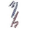
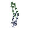
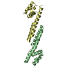
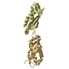
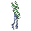
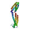
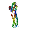
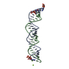
| Title | Structure of the Fluorescence Recovery Protein from Synechocystis sp PCC 6803, R60K mutant | ||||||
 Components Components | Slr1964 protein | ||||||
 Keywords Keywords | PROTEIN BINDING / photoprotection / non-photochemical quenching | ||||||
| Function / homology | Fluorescence recovery protein / : / Photoprotection regulator fluorescence recovery protein / plasma membrane-derived thylakoid membrane / Fluorescence recovery protein Function and homology information Function and homology information | ||||||
| Biological species |  | ||||||
| Method |  X-RAY DIFFRACTION / SYNCHROTRON / MOLECULAR REPLACEMENT / Resolution: 3.52 Å X-RAY DIFFRACTION / SYNCHROTRON / MOLECULAR REPLACEMENT / Resolution: 3.52 Å | ||||||
 Authors Authors | Sutter, M. / Kerfeld, C.A. | ||||||
 Citation Citation | Journal: Proc.Natl.Acad.Sci.USA / Year: 2013 Title: Crystal structure of the FRP and identification of the active site for modulation of OCP-mediated photoprotection in cyanobacteria. Authors: Sutter, M. / Wilson, A. / Leverenz, R.L. / Lopez-Igual, R. / Thurotte, A. / Salmeen, A.E. / Kirilovsky, D. / Kerfeld, C.A. | ||||||
| History |
|
- Structure visualization
| Structure viewer | Molecule: MolmilJmol/JSmol |
|---|
- Downloads & links
-Download
| PDBx/mmCIF format | 4jdq.cif.gz | 123.1 KB | Display | PDBx/mmCIF format |
|---|---|---|---|---|
| PDB format | pdb4jdq.ent.gz | 99.7 KB | Display | PDB format |
| PDBx/mmJSON format | 4jdq.json.gz | Tree view | PDBx/mmJSON format | |
| Others |  Other downloads Other downloads |
-Validation report
| Arichive directory | https://data.pdbj.org/pub/pdb/validation_reports/jd/4jdqftp://data.pdbj.org/pub/pdb/validation_reports/jd/4jdq | HTTPS FTP |
|---|
-Related structure data


-Links
 PDBj
PDBj- Assembly
| Deposited unit | 
| ||||||||
|---|---|---|---|---|---|---|---|---|---|
| 1 | 
| ||||||||
| 2 | 
| ||||||||
| 3 | 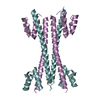
| ||||||||
| Unit cell |
|
-Components
| #1: Protein | Mass: 13186.942 Da / Num. of mol.: 6 / Mutation: R60K Source method: isolated from a genetically manipulated source Source: (gene. exp.) |
|---|
-Experimental details
-Experiment
| Experiment | Method: X-RAY DIFFRACTION / Number of used crystals: 1 |
|---|
- Sample preparation
| Crystal | Density Matthews: 2.77 Å3/Da / Density % sol: 55.59 % |
|---|---|
| Crystal grow | Temperature: 298 K / Method: vapor diffusion, sitting drop / pH: 5.5 Details: 5% PEG-3350, 100mM Na-citrate, 2% tacsimate, pH 5.5, VAPOR DIFFUSION, SITTING DROP, temperature 298K |
-Data collection
| Diffraction | Mean temperature: 100 K |
|---|---|
| Diffraction source | Source: SYNCHROTRON / Site: ALS  / Beamline: 5.0.2 / Wavelength: 1.00005 Å / Beamline: 5.0.2 / Wavelength: 1.00005 Å |
| Detector | Type: ADSC QUANTUM 315r / Detector: CCD / Date: Oct 14, 2012 |
| Radiation | Monochromator: Double-crystal, Si(111) / Protocol: SINGLE WAVELENGTH / Monochromatic (M) / Laue (L): M / Scattering type: x-ray |
| Radiation wavelength | Wavelength: 1.00005 Å / Relative weight: 1 |
| Reflection | Resolution: 3.51→39.09 Å / Num. all: 11816 / Num. obs: 11769 / % possible obs: 99.6 % / Observed criterion σ(F): 2 / Observed criterion σ(I): 2 |
| Reflection shell | Resolution: 3.51→3.7 Å / Redundancy: 13.4 % / Rmerge(I) obs: 0.01539 / Mean I/σ(I) obs: 1.6 / Num. unique all: 1625 / % possible all: 98 |
- Processing
| Software |
| ||||||||||||||||||||||||||||||||||||||||||||||||||||||||||||||||||||||||||||||||||||||||||||||||||||||||||||||||
|---|---|---|---|---|---|---|---|---|---|---|---|---|---|---|---|---|---|---|---|---|---|---|---|---|---|---|---|---|---|---|---|---|---|---|---|---|---|---|---|---|---|---|---|---|---|---|---|---|---|---|---|---|---|---|---|---|---|---|---|---|---|---|---|---|---|---|---|---|---|---|---|---|---|---|---|---|---|---|---|---|---|---|---|---|---|---|---|---|---|---|---|---|---|---|---|---|---|---|---|---|---|---|---|---|---|---|---|---|---|---|---|---|---|
| Refinement | Method to determine structure: MOLECULAR REPLACEMENT / Resolution: 3.52→39.089 Å / SU ML: 0.49 / σ(F): 1.33 / Phase error: 28.21 / Stereochemistry target values: ML
| ||||||||||||||||||||||||||||||||||||||||||||||||||||||||||||||||||||||||||||||||||||||||||||||||||||||||||||||||
| Solvent computation | Shrinkage radii: 0.9 Å / VDW probe radii: 1.11 Å / Solvent model: FLAT BULK SOLVENT MODEL | ||||||||||||||||||||||||||||||||||||||||||||||||||||||||||||||||||||||||||||||||||||||||||||||||||||||||||||||||
| Refinement step | Cycle: LAST / Resolution: 3.52→39.089 Å
| ||||||||||||||||||||||||||||||||||||||||||||||||||||||||||||||||||||||||||||||||||||||||||||||||||||||||||||||||
| Refine LS restraints |
| ||||||||||||||||||||||||||||||||||||||||||||||||||||||||||||||||||||||||||||||||||||||||||||||||||||||||||||||||
| LS refinement shell |
|
