 Movie
Movie Controller


+ Open data

- Basic information
| Entry | Database: PDB / ID: 5tvd | ||||||
|---|---|---|---|---|---|---|---|
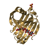
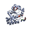
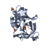
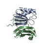
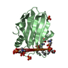
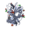
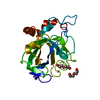
| Title | Crystal structure of Tm16 | ||||||
 Components Components | Tm16 | ||||||
 Keywords Keywords | UNKNOWN FUNCTION / phosphatidylethanolamine-binding protein | ||||||
| Function / homology |  Function and homology information Function and homology informationPhosphatidylethanolamine-binding, conserved site / Phosphatidylethanolamine-binding protein family signature. / Phosphatidylethanolamine-binding Protein / PEBP-like / Phosphatidylethanolamine-binding protein, eukaryotic / Phosphatidylethanolamine-binding protein / Phosphatidylethanolamine-binding protein / PEBP-like superfamily / Alpha-Beta Complex / Alpha Beta Similarity search - Domain/homology | ||||||
| Biological species |  Trichuris muris (invertebrata) Trichuris muris (invertebrata) | ||||||
| Method |  X-RAY DIFFRACTION / MOLECULAR REPLACEMENT / Resolution: 1.734 Å X-RAY DIFFRACTION / MOLECULAR REPLACEMENT / Resolution: 1.734 Å | ||||||
 Authors Authors | Asojo, O.A. | ||||||
 Citation Citation | Journal: J Parasitol Res / Year: 2017 Title: Identification, Characterization, and Structure of Tm16 from Trichuris muris. Authors: Liu, Z. / Kelleher, A. / Tabb, S. / Wei, J. / Pollet, J. / Hotez, P.J. / Bottazzi, M.E. / Zhan, B. / Asojo, O.A. | ||||||
| History |
|
- Structure visualization
| Structure viewer | Molecule: MolmilJmol/JSmol |
|---|
- Downloads & links
-Download
| PDBx/mmCIF format | 5tvd.cif.gz | 55 KB | Display | PDBx/mmCIF format |
|---|---|---|---|---|
| PDB format | pdb5tvd.ent.gz | 37.6 KB | Display | PDB format |
| PDBx/mmJSON format | 5tvd.json.gz | Tree view | PDBx/mmJSON format | |
| Others |  Other downloads Other downloads |
-Validation report
| Summary document | 5tvd_validation.pdf.gz | 417.6 KB | Display | wwPDB validaton report |
|---|---|---|---|---|
| Full document | 5tvd_full_validation.pdf.gz | 417.7 KB | Display | |
| Data in XML | 5tvd_validation.xml.gz | 11.3 KB | Display | |
| Data in CIF | 5tvd_validation.cif.gz | 16.6 KB | Display | |
| Arichive directory | https://data.pdbj.org/pub/pdb/validation_reports/tv/5tvdftp://data.pdbj.org/pub/pdb/validation_reports/tv/5tvd | HTTPS FTP |
-Related structure data
| Related structure data | 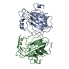 1bd9S S: Starting model for refinement |
|---|---|
| Similar structure data |


-Links
 PDBj
PDBj- Assembly
| Deposited unit | 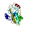
| |||||||||
|---|---|---|---|---|---|---|---|---|---|---|
| 1 |
| |||||||||
| Unit cell |
| |||||||||
| Components on special symmetry positions |
|
-Components
| #1: Protein | Mass: 21371.855 Da / Num. of mol.: 1 / Fragment: UNP residues 25-211 Source method: isolated from a genetically manipulated source Source: (gene. exp.) Trichuris muris (invertebrata) / Production host:  Pichia (fungus) / References: UniProt: A0A0N5DEJ7 Pichia (fungus) / References: UniProt: A0A0N5DEJ7 |
|---|---|
| #2: Water | ChemComp-HOH /  Mass: 18.015 Da / Num. of mol.: 244 / Source method: isolated from a natural source / Formula: H2O Mass: 18.015 Da / Num. of mol.: 244 / Source method: isolated from a natural source / Formula: H2O |
-Experimental details
-Experiment
| Experiment | Method: X-RAY DIFFRACTION / Number of used crystals: 1 |
|---|
- Sample preparation
| Crystal | Density Matthews: 2.02 Å3/Da / Density % sol: 38.98 % |
|---|---|
| Crystal grow | Temperature: 288 K / Method: vapor diffusion, sitting drop / pH: 7.5 Details: 22 mg/ml Tm16 in 5mM Bis(2-hydroxyethyl)aminotris(hydroxymethyl)methane pH 6.5 Buffer 0.1 M HEPES pH 7.5, 10% (v/v) isopropanol, 20% (w/v) PEG 4000 |
-Data collection
| Diffraction | Mean temperature: 100 K |
|---|---|
| Diffraction source | Source: ROTATING ANODE / Type: RIGAKU FR-E+ SUPERBRIGHT / Wavelength: 1.5418 Å |
| Detector | Type: RIGAKU RAXIS HR / Detector: IMAGE PLATE / Date: Aug 16, 2016 |
| Radiation | Protocol: SINGLE WAVELENGTH / Monochromatic (M) / Laue (L): M / Scattering type: x-ray |
| Radiation wavelength | Wavelength: 1.5418 Å / Relative weight: 1 |
| Reflection | Resolution: 1.734→19.93 Å / Num. obs: 17974 / % possible obs: 99.25 % / Redundancy: 1.8 % / Biso Wilson estimate: 16.31 Å2 / CC1/2: 0.995 / Rmerge(I) obs: 0.04017 / Net I/av σ(I): 10.05 / Net I/σ(I): 10.05 |
| Reflection shell | Resolution: 1.734→1.796 Å / Redundancy: 1.7 % / Rmerge(I) obs: 0.1777 / Mean I/σ(I) obs: 3.58 / CC1/2: 0.895 / % possible all: 98.2 |
- Processing
| Software |
| ||||||||||||||||||||||||||||||||||||||||||||||||||||||||
|---|---|---|---|---|---|---|---|---|---|---|---|---|---|---|---|---|---|---|---|---|---|---|---|---|---|---|---|---|---|---|---|---|---|---|---|---|---|---|---|---|---|---|---|---|---|---|---|---|---|---|---|---|---|---|---|---|---|
| Refinement | Method to determine structure: MOLECULAR REPLACEMENT Starting model: 1BD9 Resolution: 1.734→19.928 Å / SU ML: 0.19 / Cross valid method: FREE R-VALUE / σ(F): 1.34 / Phase error: 19.95
| ||||||||||||||||||||||||||||||||||||||||||||||||||||||||
| Solvent computation | Shrinkage radii: 0.9 Å / VDW probe radii: 1.11 Å | ||||||||||||||||||||||||||||||||||||||||||||||||||||||||
| Refinement step | Cycle: LAST / Resolution: 1.734→19.928 Å
| ||||||||||||||||||||||||||||||||||||||||||||||||||||||||
| Refine LS restraints |
| ||||||||||||||||||||||||||||||||||||||||||||||||||||||||
| LS refinement shell |
|
