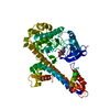
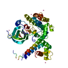
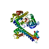
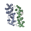
non-specific serine/threonine protein kinase / protein serine/threonine kinase activity / calcium ion binding / ATP binding Similarity search - Function
Resolution: 2.05→31.71 Å / Cor.coef. Fo:Fc: 0.9489 / Cor.coef. Fo:Fc free: 0.9368 / SU R Cruickshank DPI: 0.662 / Cross valid method: THROUGHOUT / σ(F): 0 / SU R Blow DPI: 0.199 / SU Rfree Blow DPI: 0.16 / SU Rfree Cruickshank DPI: 0.163 Details: THIS STRUCTURE MODEL WAS REFINED AND ADJUSTED USING 3KU2, A STRUCTURE MODEL OF THE SAME PROTEIN COMPLEXED WITH ANP, AS A TEMPLATE. THERE ARE GAPS IN THE PROTEIN CHAIN AND ALSO POORLY ...Details: THIS STRUCTURE MODEL WAS REFINED AND ADJUSTED USING 3KU2, A STRUCTURE MODEL OF THE SAME PROTEIN COMPLEXED WITH ANP, AS A TEMPLATE. THERE ARE GAPS IN THE PROTEIN CHAIN AND ALSO POORLY RESOLVED LOOPS WHICH ARE EASY TO MISINTERPRET. WHOEVER ANALYZES THE STRUCTURE NEEDS TO CONSIDER THE RISK OF OUT-OF-REGISTER ERRORS UNDER THESE CIRCUMSTANCES, AND IS RECOMMENDED TO REFER TO THE ELECTRON DENSITY MAP FOR MODEL CREDIBILITY.
Rfactor
Num. reflection
% reflection
Selection details
Rfree
0.2245
1377
4.94 %
RANDOM
Rwork
0.1986
-
-
-
obs
0.1999
27872
98.42 %
-
Displacement parameters
Biso mean: 66.49 Å2
Baniso -1
Baniso -2
Baniso -3
1-
5.5638 Å2
0 Å2
1.0551 Å2
2-
-
4.4809 Å2
0 Å2
3-
-
-
-10.0447 Å2
Refine analyze
Luzzati coordinate error obs: 0.292 Å
Refinement step
Cycle: LAST / Resolution: 2.05→31.71 Å
Protein
Nucleic acid
Ligand
Solvent
Total
Num. atoms
3334
0
23
113
3470
Refine LS restraints
Refine-ID
Type
Dev ideal
Number
Restraint function
Weight
X-RAY DIFFRACTION
t_bond_d
0.008
6577
HARMONIC
2
X-RAY DIFFRACTION
t_angle_deg
0.86
11829
HARMONIC
2
X-RAY DIFFRACTION
t_dihedral_angle_d
1455
SINUSOIDAL
2
X-RAY DIFFRACTION
t_incorr_chiral_ct
X-RAY DIFFRACTION
t_pseud_angle
X-RAY DIFFRACTION
t_trig_c_planes
80
HARMONIC
2
X-RAY DIFFRACTION
t_gen_planes
1023
HARMONIC
5
X-RAY DIFFRACTION
t_it
6577
HARMONIC
20
X-RAY DIFFRACTION
t_nbd
X-RAY DIFFRACTION
t_omega_torsion
2.56
X-RAY DIFFRACTION
t_other_torsion
14.95
X-RAY DIFFRACTION
t_improper_torsion
X-RAY DIFFRACTION
t_chiral_improper_torsion
460
SEMIHARMONIC
5
X-RAY DIFFRACTION
t_sum_occupancies
X-RAY DIFFRACTION
t_utility_distance
X-RAY DIFFRACTION
t_utility_angle
X-RAY DIFFRACTION
t_utility_torsion
X-RAY DIFFRACTION
t_ideal_dist_contact
7029
SEMIHARMONIC
4
LS refinement shell
Resolution: 2.05→2.13 Å / Total num. of bins used: 14
Rfactor
Num. reflection
% reflection
Rfree
0.2225
132
4.65 %
Rwork
0.2231
2706
-
all
0.2231
2838
-
obs
-
-
94.93 %
Refinement TLS params.
Method: refined / Refine-ID: X-RAY DIFFRACTION
ID
L11 (°2)
L12 (°2)
L13 (°2)
L22 (°2)
L23 (°2)
L33 (°2)
S11 (Å °)
S12 (Å °)
S13 (Å °)
S21 (Å °)
S22 (Å °)
S23 (Å °)
S31 (Å °)
S32 (Å °)
S33 (Å °)
T11 (Å2)
T12 (Å2)
T13 (Å2)
T22 (Å2)
T23 (Å2)
T33 (Å2)
Origin x (Å)
Origin y (Å)
Origin z (Å)
1
4.2646
1.1332
-0.3067
4.1582
0.8226
3.8609
-0.0816
-0.1389
0.2735
0.1693
0.0619
-0.1639
-0.2528
0.1239
0.0197
-0.079
-0.0387
-0.0797
-0.1919
0.0809
0.0288
3.6755
-9.6241
41.722
2
1.5802
0.118
0.4542
2.839
-2.1536
3.3322
-0.028
0.012
-0.259
-0.0575
-0.1595
-0.2423
0.2263
0.151
0.1875
-0.1908
0.0203
0.0521
-0.2161
0.0331
0.0349
-3.5492
-7.8138
25.9614
3
1.9509
0.1436
0.8077
1.7146
-0.4513
2.1765
-0.1006
0.373
-0.1129
-0.3386
0.0021
0.0327
0.2066
0.1824
0.0985
-0.1669
0.0436
0.0728
-0.1223
0.0001
-0.1153
-1.016
1.6695
13.3722
4
2.7248
-1.4777
-0.0267
0
1.2083
4.644
0.1122
-0.0405
0.245
0.2766
-0.1206
-0.1845
-0.1946
0.0086
0.0085
0.3582
-0.1561
-0.0875
-0.2768
-0.036
-0.2327
21.8557
6.8921
51.4712
5
7.0171
-2.0423
-2.2805
3.8797
1.1014
3.8767
0.1283
0.0326
0.361
0.1044
0.1212
0.0687
-0.2922
-0.2381
-0.2495
-0.1695
0.0337
0.0056
-0.1031
0.0897
-0.021
18.3438
15.5913
21.5737
Refinement TLS group
ID
Refine-ID
Refine TLS-ID
Selection details
1
X-RAY DIFFRACTION
1
{ A|44 - 75 }
2
X-RAY DIFFRACTION
2
{ A|76 - 213 }
3
X-RAY DIFFRACTION
3
{ A|214 - 364 }
4
X-RAY DIFFRACTION
4
{ A|366 - 440 }
5
X-RAY DIFFRACTION
5
{ A|441 - 507 }
+
About Yorodumi
-
News
-
Feb 9, 2022. New format data for meta-information of EMDB entries
New format data for meta-information of EMDB entries
Version 3 of the EMDB header file is now the official format.
The previous official version 1.9 will be removed from the archive.
In the structure databanks used in Yorodumi, some data are registered as the other names, "COVID-19 virus" and "2019-nCoV". Here are the details of the virus and the list of structure data.
Jan 31, 2019. EMDB accession codes are about to change! (news from PDBe EMDB page)
EMDB accession codes are about to change! (news from PDBe EMDB page)
The allocation of 4 digits for EMDB accession codes will soon come to an end. Whilst these codes will remain in use, new EMDB accession codes will include an additional digit and will expand incrementally as the available range of codes is exhausted. The current 4-digit format prefixed with “EMD-” (i.e. EMD-XXXX) will advance to a 5-digit format (i.e. EMD-XXXXX), and so on. It is currently estimated that the 4-digit codes will be depleted around Spring 2019, at which point the 5-digit format will come into force.
The EM Navigator/Yorodumi systems omit the EMD- prefix.
Related info.:Q: What is EMD? / ID/Accession-code notation in Yorodumi/EM Navigator
Yorodumi is a browser for structure data from EMDB, PDB, SASBDB, etc.
This page is also the successor to EM Navigator detail page, and also detail information page/front-end page for Omokage search.
The word "yorodu" (or yorozu) is an old Japanese word meaning "ten thousand". "mi" (miru) is to see.
Related info.:EMDB / PDB / SASBDB / Comparison of 3 databanks / Yorodumi Search / Aug 31, 2016. New EM Navigator & Yorodumi / Yorodumi Papers / Jmol/JSmol / Function and homology information / Changes in new EM Navigator and Yorodumi
 Movie
Movie Controller

 Yorodumi
 Open data
 Basic information
 Components
 Keywords
 Function and homology information
 X-RAY DIFFRACTION /
  Authors
 Citation
 Structure visualization
 Downloads & links
 Other downloads
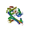


 PDBj
PDBj Assembly

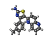
 Mass: 318.396 Da / Num. of mol.: 1 / Source method: obtained synthetically / Formula: C18H14N4S
 Mass: 18.015 Da / Num. of mol.: 113 / Source method: isolated from a natural source / Formula: H2O
 Sample preparation
 / Beamline: 24-ID-E / Wavelength: 0.97921 Å
 Processing