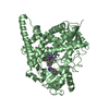
non-specific serine/threonine protein kinase / protein serine/threonine kinase activity / calcium ion binding / ATP binding Similarity search - Function
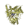
Resolution: 2.4→31.644 Å / SU ML: 0.4 / Cross valid method: FREE R-VALUE / σ(F): 1.35 / Phase error: 35.58 / Stereochemistry target values: ML Details: This structure model was refined and adjusted using 3KU2, a structure model of the same protein complexed with ANP, as a template. It contains disjoint segments around residues 358, 377 and ...Details: This structure model was refined and adjusted using 3KU2, a structure model of the same protein complexed with ANP, as a template. It contains disjoint segments around residues 358, 377 and 405, and the main chain torsion angles are remarkably poor in these residues. There are also other gaps in the protein chain and poorly resolved loops which are easy to misinterpret. Whoever analyzes the structure needs to consider the risk of out-of-register errors under these circumstances, and is recommended to refer to the electron density map for model credibility.
Rfactor
Num. reflection
% reflection
Rfree
0.299
850
4.93 %
Rwork
0.2303
16400
-
obs
0.2338
17250
98.48 %
Solvent computation
Shrinkage radii: 0.9 Å / VDW probe radii: 1.11 Å / Solvent model: FLAT BULK SOLVENT MODEL
In the structure databanks used in Yorodumi, some data are registered as the other names, "COVID-19 virus" and "2019-nCoV". Here are the details of the virus and the list of structure data.
Jan 31, 2019. EMDB accession codes are about to change! (news from PDBe EMDB page)
EMDB accession codes are about to change! (news from PDBe EMDB page)
The allocation of 4 digits for EMDB accession codes will soon come to an end. Whilst these codes will remain in use, new EMDB accession codes will include an additional digit and will expand incrementally as the available range of codes is exhausted. The current 4-digit format prefixed with “EMD-” (i.e. EMD-XXXX) will advance to a 5-digit format (i.e. EMD-XXXXX), and so on. It is currently estimated that the 4-digit codes will be depleted around Spring 2019, at which point the 5-digit format will come into force.
The EM Navigator/Yorodumi systems omit the EMD- prefix.
Related info.:Q: What is EMD? / ID/Accession-code notation in Yorodumi/EM Navigator
Yorodumi is a browser for structure data from EMDB, PDB, SASBDB, etc.
This page is also the successor to EM Navigator detail page, and also detail information page/front-end page for Omokage search.
The word "yorodu" (or yorozu) is an old Japanese word meaning "ten thousand". "mi" (miru) is to see.
Related info.:EMDB / PDB / SASBDB / Comparison of 3 databanks / Yorodumi Search / Aug 31, 2016. New EM Navigator & Yorodumi / Yorodumi Papers / Jmol/JSmol / Function and homology information / Changes in new EM Navigator and Yorodumi
 Movie
Movie Controller

 Yorodumi
 Open data
 Basic information
 Components
 Keywords
 Function and homology information
 X-RAY DIFFRACTION /
  Authors
 Citation
 Structure visualization
 Downloads & links
 Other downloads
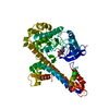


 PDBj
PDBj Assembly


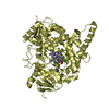
 Mass: 338.814 Da / Num. of mol.: 1 / Source method: obtained synthetically / Formula: C17H11ClN4S
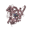
 Num. of mol.: 7 / Source method: obtained synthetically
 Mass: 18.015 Da / Num. of mol.: 3 / Source method: isolated from a natural source / Formula: H2O
 Sample preparation
 / Beamline: 24-ID-E / Wavelength: 0.97921 Å
 Processing