 Movie
Movie Controller

[English] 日本語
 Yorodumi
- PDB-5t5n: Calcium-activated chloride channel bestrophin-1 (BEST1), triple m... -
+ Open data

- Basic information
| Entry | Database: PDB / ID: 5t5n | ||||||
|---|---|---|---|---|---|---|---|
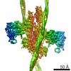
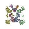
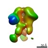
| Title | Calcium-activated chloride channel bestrophin-1 (BEST1), triple mutant: I76A, F80A, F84A; in complex with an Fab antibody fragment, chloride, and calcium | ||||||
 Components Components |
| ||||||
 Keywords Keywords | MEMBRANE PROTEIN / ion channel / anion / chloride / calcium-activated / eukaryotic membrane protein / CaCC | ||||||
| Function / homology |  Function and homology information Function and homology informationStimuli-sensing channels / chloride channel activity / membrane / metal ion binding Similarity search - Function | ||||||
| Biological species |   | ||||||
| Method |  X-RAY DIFFRACTION / SYNCHROTRON / MOLECULAR REPLACEMENT / Resolution: 3.1 Å X-RAY DIFFRACTION / SYNCHROTRON / MOLECULAR REPLACEMENT / Resolution: 3.1 Å | ||||||
 Authors Authors | Long, S.B. / Vaisey, G. | ||||||
| Funding support |  United States, 1items United States, 1items
| ||||||
 Citation Citation | Journal: Proc. Natl. Acad. Sci. U.S.A. / Year: 2016 Title: Distinct regions that control ion selectivity and calcium-dependent activation in the bestrophin ion channel. Authors: Vaisey, G. / Miller, A.N. / Long, S.B. | ||||||
| History |
|
- Structure visualization
| Structure viewer | Molecule: MolmilJmol/JSmol |
|---|
- Downloads & links
-Download
| PDBx/mmCIF format | 5t5n.cif.gz | 756 KB | Display | PDBx/mmCIF format |
|---|---|---|---|---|
| PDB format | pdb5t5n.ent.gz | 620.7 KB | Display | PDB format |
| PDBx/mmJSON format | 5t5n.json.gz | Tree view | PDBx/mmJSON format | |
| Others |  Other downloads Other downloads |
-Validation report
| Arichive directory | https://data.pdbj.org/pub/pdb/validation_reports/t5/5t5nftp://data.pdbj.org/pub/pdb/validation_reports/t5/5t5n | HTTPS FTP |
|---|
-Related structure data
| Related structure data | 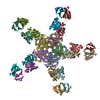 4rdqS S: Starting model for refinement |
|---|---|
| Similar structure data |


-Links
 PDBj
PDBj

- Assembly
| Deposited unit | 
| ||||||||
|---|---|---|---|---|---|---|---|---|---|
| 1 |
| ||||||||
| Unit cell |
|
-Components
-Protein , 1 types, 5 molecules ABCDE
| #1: Protein | Mass: 47420.070 Da / Num. of mol.: 5 / Mutation: I76A, F80A, F84A Source method: isolated from a genetically manipulated source Source: (gene. exp.)  Pichia (fungus) / References: UniProt: E1C3A0 Pichia (fungus) / References: UniProt: E1C3A0 |
|---|
-Antibody , 2 types, 10 molecules FHJLNGIKMO
| #2: Antibody | Mass: 23339.707 Da / Num. of mol.: 5 / Source method: isolated from a natural source / Source: (natural) #3: Antibody | Mass: 23638.525 Da / Num. of mol.: 5 / Source method: isolated from a natural source / Source: (natural) |
|---|
-Non-polymers , 5 types, 40 molecules 


| #4: Chemical | ChemComp-CL /  Mass: 35.453 Da / Num. of mol.: 15 / Source method: obtained synthetically / Formula: Cl Mass: 35.453 Da / Num. of mol.: 15 / Source method: obtained synthetically / Formula: Cl#5: Chemical | ChemComp-K /  Mass: 39.098 Da / Num. of mol.: 5 / Source method: obtained synthetically / Formula: K Mass: 39.098 Da / Num. of mol.: 5 / Source method: obtained synthetically / Formula: K#6: Chemical | ChemComp-GOL /  Mass: 92.094 Da / Num. of mol.: 5 / Source method: obtained synthetically / Formula: C3H8O3 Mass: 92.094 Da / Num. of mol.: 5 / Source method: obtained synthetically / Formula: C3H8O3#7: Chemical | ChemComp-CA /  Mass: 40.078 Da / Num. of mol.: 5 / Source method: obtained synthetically / Formula: Ca Mass: 40.078 Da / Num. of mol.: 5 / Source method: obtained synthetically / Formula: Ca#8: Water | ChemComp-HOH / | Mass: 18.015 Da / Num. of mol.: 10 / Source method: isolated from a natural source / Formula: H2O |
|---|
-Details
| Has protein modification | Y |
|---|
-Experimental details
-Experiment
| Experiment | Method: X-RAY DIFFRACTION / Number of used crystals: 1 |
|---|
- Sample preparation
| Crystal | Density Matthews: 4.36 Å3/Da / Density % sol: 71.77 % |
|---|---|
| Crystal grow | Temperature: 293 K / Method: vapor diffusion, hanging drop / pH: 4 Details: 6% Peg 4000 50 mM sodium acetate pH 4.0 20% glycerol 75 mM NaCl 75 mM KCl 10 mM Tris-HCl pH 7.5 0.5 mM Cymal-6-NG (Anatrace) |
-Data collection
| Diffraction | Mean temperature: 100 K |
|---|---|
| Diffraction source | Source: SYNCHROTRON / Site: APS / Beamline: 23-ID-D / Wavelength: 1.033 Å |
| Detector | Type: DECTRIS PILATUS3 6M / Detector: PIXEL / Date: Dec 1, 2014 |
| Radiation | Monochromator: Si(111) / Protocol: SINGLE WAVELENGTH / Monochromatic (M) / Laue (L): M / Scattering type: x-ray |
| Radiation wavelength | Wavelength: 1.033 Å / Relative weight: 1 |
| Reflection | Resolution: 3.1→38 Å / Num. obs: 145877 / % possible obs: 99.9 % / Observed criterion σ(I): -3 / Redundancy: 9.5 % / Biso Wilson estimate: 82 Å2 / Net I/σ(I): 9.7 |
| Reflection shell | Resolution: 3.1→3.15 Å / Redundancy: 9.7 % / Mean I/σ(I) obs: 1.2 / CC1/2: 0.604 / % possible all: 99.9 |
- Processing
| Software |
| ||||||||||||||||||||
|---|---|---|---|---|---|---|---|---|---|---|---|---|---|---|---|---|---|---|---|---|---|
| Refinement | Method to determine structure: MOLECULAR REPLACEMENT Starting model: PDB ID 4RDQ Resolution: 3.1→37.952 Å / Cross valid method: THROUGHOUT / Phase error: 31.19
| ||||||||||||||||||||
| Displacement parameters | Biso mean: 101.2 Å2 | ||||||||||||||||||||
| Refinement step | Cycle: LAST / Resolution: 3.1→37.952 Å
| ||||||||||||||||||||
| LS refinement shell | Resolution: 3.1→3.211 Å
|
