| Entry | Database: PDB / ID: 5t3j
|
|---|
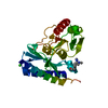
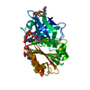
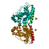
| Title | Histidinol Phosphate Phosphatase(HPP) soaked with selenourea for 10 min |
|---|
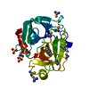
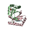
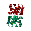
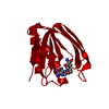
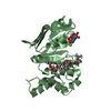
 Components Components | Inositol monophosphatase |
|---|
 Keywords Keywords | HYDROLASE / Histidinol Phosphate Phosphatase(HPP) / selenourea |
|---|
| Function / homology |  Function and homology information Function and homology information
Histidinol-phosphate phosphatase, putative, inositol monophosphatase / : / Inositol monophosphatase, metal-binding site / Inositol monophosphatase family signature 1. / Inositol monophosphatase-like / Inositol monophosphatase family / D-Maltodextrin-Binding Protein; domain 2 - #80 / Fructose-1,6-Bisphosphatase, subunit A, domain 1 / Fructose-1,6-Bisphosphatase; Chain A, domain 1 / D-Maltodextrin-Binding Protein; domain 2 ...Histidinol-phosphate phosphatase, putative, inositol monophosphatase / : / Inositol monophosphatase, metal-binding site / Inositol monophosphatase family signature 1. / Inositol monophosphatase-like / Inositol monophosphatase family / D-Maltodextrin-Binding Protein; domain 2 - #80 / Fructose-1,6-Bisphosphatase, subunit A, domain 1 / Fructose-1,6-Bisphosphatase; Chain A, domain 1 / D-Maltodextrin-Binding Protein; domain 2 / 2-Layer Sandwich / 3-Layer(aba) Sandwich / Alpha BetaSimilarity search - Domain/homology |
|---|
| Biological species |   Medicago truncatula (barrel medic) Medicago truncatula (barrel medic) |
|---|
| Method |  X-RAY DIFFRACTION / SYNCHROTRON / SAD / Resolution: 2.55 Å X-RAY DIFFRACTION / SYNCHROTRON / SAD / Resolution: 2.55 Å |
|---|
 Authors Authors | Luo, Z. / Dauter, Z. |
|---|
| Funding support |  United States, 1items United States, 1items | Organization | Grant number | Country |
|---|
| National Institutes of Health/National Cancer Institute (NIH/NCI) | | United States |
|
|---|
 Citation Citation | Journal: Sci Rep / Year: 2016
Title: Selenourea: a convenient phasing vehicle for macromolecular X-ray crystal structures.
Authors: Luo, Z. |
|---|
| History | | Deposition | Aug 25, 2016 | Deposition site: RCSB / Processing site: RCSB |
|---|
| Revision 1.0 | Nov 30, 2016 | Provider: repository / Type: Initial release |
|---|
| Revision 1.1 | Mar 15, 2017 | Group: Refinement description |
|---|
| Revision 1.2 | Sep 27, 2017 | Group: Author supporting evidence / Category: pdbx_audit_support / Item: _pdbx_audit_support.funding_organization |
|---|
| Revision 1.3 | Dec 4, 2019 | Group: Author supporting evidence / Derived calculations / Category: pdbx_audit_support / pdbx_struct_special_symmetry / Item: _pdbx_audit_support.funding_organization |
|---|
| Revision 1.4 | Mar 6, 2024 | Group: Data collection / Database references / Category: chem_comp_atom / chem_comp_bond / database_2
Item: _database_2.pdbx_DOI / _database_2.pdbx_database_accession |
|---|
|
|---|
 Movie
Movie Controller

 Yorodumi
 Open data
 Basic information
 Structure visualization
 Downloads & links
 Other downloads
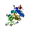


 PDBj
PDBj Assembly
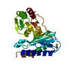


 Mass: 123.016 Da / Num. of mol.: 1 / Source method: obtained synthetically / Formula: CH4N2Se
 Mass: 94.971 Da / Num. of mol.: 1 / Source method: obtained synthetically / Formula: PO4
 Mass: 18.015 Da / Num. of mol.: 79 / Source method: isolated from a natural source / Formula: H2O
 Sample preparation
 Processing