 Movie
Movie Controller

[English] 日本語
 Yorodumi
- PDB-1oii: Crystal structure of the alkylsulfatase AtsK, a non-heme Fe(II) a... -
+ Open data

- Basic information
| Entry | Database: PDB / ID: 1oii | ||||||
|---|---|---|---|---|---|---|---|
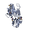
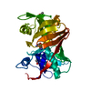
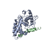
| Title | Crystal structure of the alkylsulfatase AtsK, a non-heme Fe(II) alphaketoglutarate dependent Dioxygenase in complex with iron and alphaketoglutarate | ||||||
 Components Components | PUTATIVE ALKYLSULFATASE ATSK | ||||||
 Keywords Keywords | OXIDOREDUCTASE / JELLY ROLL | ||||||
| Function / homology |  Function and homology information Function and homology informationalkyl sulfatase / 2-oxoglutarate-dependent dioxygenase activity / metal ion binding / cytoplasm Similarity search - Function | ||||||
| Biological species |  PSEUDOMONAS PUTIDA (bacteria) PSEUDOMONAS PUTIDA (bacteria) | ||||||
| Method |  X-RAY DIFFRACTION / SYNCHROTRON / MOLECULAR REPLACEMENT / Resolution: 2.19 Å X-RAY DIFFRACTION / SYNCHROTRON / MOLECULAR REPLACEMENT / Resolution: 2.19 Å | ||||||
 Authors Authors | Mueller, I. / Kahnert, A. / Pape, T. / Dierks, T. / Meyer-Klauke, W. / Kertesz, M.A. / Uson, I. | ||||||
 Citation Citation | Journal: Biochemistry / Year: 2004 Title: Crystal Structure of the Alkylsulfatase Atsk: Insights Into the Catalytic Mechanism of the Fe(II) Alpha-Ketoglutarate-Dependent Dioxygenase Superfamily Authors: Mueller, I. / Kahnert, A. / Pape, T. / Sheldrick, G.M. / Meyer-Klaucke, W. / Dierks, T. / Kertesz, M.A. / Uson, I. | ||||||
| History |
|
- Structure visualization
| Structure viewer | Molecule: MolmilJmol/JSmol |
|---|
- Downloads & links
-Download
| PDBx/mmCIF format | 1oii.cif.gz | 215 KB | Display | PDBx/mmCIF format |
|---|---|---|---|---|
| PDB format | pdb1oii.ent.gz | 171.8 KB | Display | PDB format |
| PDBx/mmJSON format | 1oii.json.gz | Tree view | PDBx/mmJSON format | |
| Others |  Other downloads Other downloads |
-Validation report
| Arichive directory | https://data.pdbj.org/pub/pdb/validation_reports/oi/1oiiftp://data.pdbj.org/pub/pdb/validation_reports/oi/1oii | HTTPS FTP |
|---|
-Related structure data
| Related structure data | 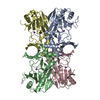 1oihSC 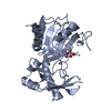 1oijC 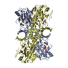 1oikC S: Starting model for refinement C: citing same article ( |
|---|---|
| Similar structure data |


-Links
 PDBj
PDBj


- Assembly
| Deposited unit | 
| |||||||||||||||||||||||||||||||||||||||||||||||||||||||||||||||||||||||||||||||||||||||||||||||||||||||||||||
|---|---|---|---|---|---|---|---|---|---|---|---|---|---|---|---|---|---|---|---|---|---|---|---|---|---|---|---|---|---|---|---|---|---|---|---|---|---|---|---|---|---|---|---|---|---|---|---|---|---|---|---|---|---|---|---|---|---|---|---|---|---|---|---|---|---|---|---|---|---|---|---|---|---|---|---|---|---|---|---|---|---|---|---|---|---|---|---|---|---|---|---|---|---|---|---|---|---|---|---|---|---|---|---|---|---|---|---|---|---|---|
| 1 | 
| |||||||||||||||||||||||||||||||||||||||||||||||||||||||||||||||||||||||||||||||||||||||||||||||||||||||||||||
| 2 | 
| |||||||||||||||||||||||||||||||||||||||||||||||||||||||||||||||||||||||||||||||||||||||||||||||||||||||||||||
| 3 | 
| |||||||||||||||||||||||||||||||||||||||||||||||||||||||||||||||||||||||||||||||||||||||||||||||||||||||||||||
| 4 | 
| |||||||||||||||||||||||||||||||||||||||||||||||||||||||||||||||||||||||||||||||||||||||||||||||||||||||||||||
| Unit cell |
| |||||||||||||||||||||||||||||||||||||||||||||||||||||||||||||||||||||||||||||||||||||||||||||||||||||||||||||
| Noncrystallographic symmetry (NCS) | NCS domain:
NCS domain segments:
NCS oper:
|
-Components
| #1: Protein | Mass: 33248.402 Da / Num. of mol.: 4 / Source method: isolated from a natural source / Details: DSM 6884 / Source: (natural) PSEUDOMONAS PUTIDA (bacteria) / Strain: S-313 / References: UniProt: Q9WWU5#2: Chemical | ChemComp-FE2 /   Mass: 55.845 Da / Num. of mol.: 4 / Source method: obtained synthetically / Formula: Fe Mass: 55.845 Da / Num. of mol.: 4 / Source method: obtained synthetically / Formula: Fe#3: Chemical | ChemComp-AKG /   Mass: 146.098 Da / Num. of mol.: 4 / Source method: obtained synthetically / Formula: C5H6O5 Mass: 146.098 Da / Num. of mol.: 4 / Source method: obtained synthetically / Formula: C5H6O5#4: Water | ChemComp-HOH / |  Mass: 18.015 Da / Num. of mol.: 573 / Source method: isolated from a natural source / Formula: H2O Mass: 18.015 Da / Num. of mol.: 573 / Source method: isolated from a natural source / Formula: H2O |
|---|
-Experimental details
-Experiment
| Experiment | Method: X-RAY DIFFRACTION / Number of used crystals: 1 |
|---|
- Sample preparation
| Crystal | Density Matthews: 3.18 Å3/Da / Density % sol: 60 % | ||||||||||||||||||||||||||||||||||||||||||
|---|---|---|---|---|---|---|---|---|---|---|---|---|---|---|---|---|---|---|---|---|---|---|---|---|---|---|---|---|---|---|---|---|---|---|---|---|---|---|---|---|---|---|---|
| Crystal grow | pH: 5.6 Details: 50 MM NA SULFATE, 50 MM NA CITRATE PH 5.5, 10% PEG 4000, 30% GLYCEROL | ||||||||||||||||||||||||||||||||||||||||||
| Crystal grow | *PLUS Temperature: 20 ℃ / pH: 5.6 / Method: vapor diffusion, hanging drop | ||||||||||||||||||||||||||||||||||||||||||
| Components of the solutions | *PLUS
|
-Data collection
| Diffraction | Mean temperature: 100 K |
|---|---|
| Diffraction source | Source: SYNCHROTRON / Site: EMBL/DESY, HAMBURG  / Beamline: X11 / Wavelength: 0.8098 / Beamline: X11 / Wavelength: 0.8098 |
| Detector | Type: MARRESEARCH / Detector: CCD / Details: PREMIRROR, TRIANGULAR MONOCHROMATOR, BENT MIRROR |
| Radiation | Monochromator: MIRROR / Protocol: SINGLE WAVELENGTH / Monochromatic (M) / Laue (L): M / Scattering type: x-ray |
| Radiation wavelength | Wavelength: 0.8098 Å / Relative weight: 1 |
| Reflection | Resolution: 2.19→20 Å / Num. obs: 86268 / % possible obs: 97.5 % / Redundancy: 6.38 % / Rmerge(I) obs: 0.0644 / Net I/σ(I): 19.96 |
| Reflection shell | Resolution: 2.19→2.3 Å / Redundancy: 5.61 % / Rmerge(I) obs: 0.452 / Mean I/σ(I) obs: 5.1 / % possible all: 83.1 |
| Reflection | *PLUS Highest resolution: 2.19 Å / Lowest resolution: 20 Å / Num. measured all: 564785 / Rmerge(I) obs: 0.064 |
| Reflection shell | *PLUS Lowest resolution: 2.3 Å / % possible obs: 83.1 % / Rmerge(I) obs: 0.452 / Mean I/σ(I) obs: 5.1 |
- Processing
| Software |
| ||||||||||||||||||||||||||||||||||||||||||||||||||||||||||||||||||||||||||||||||||||||||||||||||||||||||||||||||||||||||||||||||||||||||||||||||||||||||||||||||||||||||||||||||||||||
|---|---|---|---|---|---|---|---|---|---|---|---|---|---|---|---|---|---|---|---|---|---|---|---|---|---|---|---|---|---|---|---|---|---|---|---|---|---|---|---|---|---|---|---|---|---|---|---|---|---|---|---|---|---|---|---|---|---|---|---|---|---|---|---|---|---|---|---|---|---|---|---|---|---|---|---|---|---|---|---|---|---|---|---|---|---|---|---|---|---|---|---|---|---|---|---|---|---|---|---|---|---|---|---|---|---|---|---|---|---|---|---|---|---|---|---|---|---|---|---|---|---|---|---|---|---|---|---|---|---|---|---|---|---|---|---|---|---|---|---|---|---|---|---|---|---|---|---|---|---|---|---|---|---|---|---|---|---|---|---|---|---|---|---|---|---|---|---|---|---|---|---|---|---|---|---|---|---|---|---|---|---|---|---|
| Refinement | Method to determine structure: MOLECULAR REPLACEMENT Starting model: PDB ENTRY 1OIH Resolution: 2.19→20 Å / Cor.coef. Fo:Fc: 0.946 / Cor.coef. Fo:Fc free: 0.934 / SU B: 4.18 / SU ML: 0.107 / Cross valid method: THROUGHOUT / ESU R: 0.179 / ESU R Free: 0.156 / Stereochemistry target values: MAXIMUM LIKELIHOOD
| ||||||||||||||||||||||||||||||||||||||||||||||||||||||||||||||||||||||||||||||||||||||||||||||||||||||||||||||||||||||||||||||||||||||||||||||||||||||||||||||||||||||||||||||||||||||
| Solvent computation | Ion probe radii: 0.8 Å / Shrinkage radii: 0.8 Å / VDW probe radii: 1.4 Å / Solvent model: BABINET MODEL WITH MASK | ||||||||||||||||||||||||||||||||||||||||||||||||||||||||||||||||||||||||||||||||||||||||||||||||||||||||||||||||||||||||||||||||||||||||||||||||||||||||||||||||||||||||||||||||||||||
| Displacement parameters | Biso mean: 33.95 Å2
| ||||||||||||||||||||||||||||||||||||||||||||||||||||||||||||||||||||||||||||||||||||||||||||||||||||||||||||||||||||||||||||||||||||||||||||||||||||||||||||||||||||||||||||||||||||||
| Refinement step | Cycle: LAST / Resolution: 2.19→20 Å
| ||||||||||||||||||||||||||||||||||||||||||||||||||||||||||||||||||||||||||||||||||||||||||||||||||||||||||||||||||||||||||||||||||||||||||||||||||||||||||||||||||||||||||||||||||||||
| Refine LS restraints |
|