 Movie
Movie Controller

[English] 日本語
 Yorodumi
- PDB-3aax: Crystal structure of probable thiosulfate sulfurtransferase cysa3... -
+ Open data

- Basic information
| Entry | Database: PDB / ID: 3aax | ||||||
|---|---|---|---|---|---|---|---|
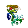
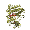
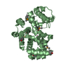
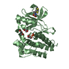
| Title | Crystal structure of probable thiosulfate sulfurtransferase cysa3 (RV3117) from Mycobacterium tuberculosis: monoclinic FORM | ||||||
 Components Components | Putative thiosulfate sulfurtransferase | ||||||
 Keywords Keywords | TRANSFERASE / M. tuberculosis / sulfurtransferase / Structural Genomics / PSI / Protein Structure Initiative / TB Structural Genomics Consortium (TBSGC) | ||||||
| Function / homology |  Function and homology information Function and homology informationthiosulfate sulfurtransferase / thiosulfate-cyanide sulfurtransferase activity / peptidoglycan-based cell wall / plasma membrane / cytosol Similarity search - Function | ||||||
| Biological species |   Mycobacterium tuberculosis (bacteria) Mycobacterium tuberculosis (bacteria) | ||||||
| Method |  X-RAY DIFFRACTION / SYNCHROTRON / MOLECULAR REPLACEMENT / Resolution: 2.5 Å X-RAY DIFFRACTION / SYNCHROTRON / MOLECULAR REPLACEMENT / Resolution: 2.5 Å | ||||||
 Authors Authors | Sankaranarayanan, R. / Witholt, S.J. / Cherney, M.M. / Garen, C.R. / Cherney, L.T. / James, M.N.G. / TB Structural Genomics Consortium (TBSGC) | ||||||
 Citation Citation | Journal: To be Published Title: The crystal structure of probable thiosulfate sulfurtransferase CysA3 (Rv3117) from Mycobacterium tuberculosis Authors: Sankaranarayanan, R. / Witholt, S.J. / Cherney, M.M. / Garen, C.R. / Cherney, L.T. / James, M.N.G. #1: Journal: Acta Crystallogr.,Sect.F / Year: 2008 Title: Expression, purification, crystallization and preliminary X-ray analysis of Rv3117, a probable thiosulfate sulfurtransferase (CysA3) from Mycobacterium tuberculosis Authors: Witholt, S.J. / Sankaranarayanan, R. / Garen, C.R. / Cherney, M.M. / Cherney, L.T. / James, M.N.G. | ||||||
| History |
|
- Structure visualization
| Structure viewer | Molecule: MolmilJmol/JSmol |
|---|
- Downloads & links
-Download
| PDBx/mmCIF format | 3aax.cif.gz | 116.1 KB | Display | PDBx/mmCIF format |
|---|---|---|---|---|
| PDB format | pdb3aax.ent.gz | 90.6 KB | Display | PDB format |
| PDBx/mmJSON format | 3aax.json.gz | Tree view | PDBx/mmJSON format | |
| Others |  Other downloads Other downloads |
-Validation report
| Arichive directory | https://data.pdbj.org/pub/pdb/validation_reports/aa/3aaxftp://data.pdbj.org/pub/pdb/validation_reports/aa/3aax | HTTPS FTP |
|---|
-Related structure data
| Related structure data |  3aayC  1uarS C: citing same article ( S: Starting model for refinement |
|---|---|
| Similar structure data | |
| Other databases |


-Links
 PDBj
PDBj- Assembly
| Deposited unit | 
| ||||||||
|---|---|---|---|---|---|---|---|---|---|
| 1 | 
| ||||||||
| 2 | 
| ||||||||
| Unit cell |
|
-Components
| #1: Protein | Mass: 31052.516 Da / Num. of mol.: 2 Source method: isolated from a genetically manipulated source Source: (gene. exp.) Mycobacterium tuberculosis (bacteria) / Strain: H37RV / Gene: CysA3 / Plasmid: pDEST-17 / Production host: References: UniProt: O05793, UniProt: P9WHF9*PLUS, thiosulfate sulfurtransferase #2: Water | ChemComp-HOH / |  Mass: 18.015 Da / Num. of mol.: 68 / Source method: isolated from a natural source / Formula: H2O Mass: 18.015 Da / Num. of mol.: 68 / Source method: isolated from a natural source / Formula: H2O |
|---|
-Experimental details
-Experiment
| Experiment | Method: X-RAY DIFFRACTION / Number of used crystals: 1 |
|---|
- Sample preparation
| Crystal | Density Matthews: 2.37 Å3/Da / Density % sol: 48.21 % |
|---|---|
| Crystal grow | Temperature: 298 K / Method: vapor diffusion, hanging drop / pH: 8.5 Details: Protein concentration 8mg/ml, 0.1M Tris HCl pH 8.5, Precipitant 0.2M MgCl2, 25% PEG 3350., VAPOR DIFFUSION, HANGING DROP, temperature 298K |
-Data collection
| Diffraction | Mean temperature: 100 K |
|---|---|
| Diffraction source | Source: SYNCHROTRON / Site: ALS  / Beamline: 8.3.1 / Wavelength: 1.11587 Å / Beamline: 8.3.1 / Wavelength: 1.11587 Å |
| Detector | Type: ADSC QUANTUM 315r / Detector: CCD / Date: Nov 30, 2007 |
| Radiation | Monochromator: Double flat crystal, Si(iii) / Protocol: SINGLE WAVELENGTH / Monochromatic (M) / Laue (L): M / Scattering type: x-ray |
| Radiation wavelength | Wavelength: 1.11587 Å / Relative weight: 1 |
| Reflection | Resolution: 2.5→45.7 Å / Num. all: 20078 / Num. obs: 20078 / % possible obs: 98.8 % / Observed criterion σ(F): 0 / Observed criterion σ(I): 0 / Redundancy: 3.6 % / Rmerge(I) obs: 0.104 / Net I/σ(I): 11.8 |
| Reflection shell | Resolution: 2.5→2.59 Å / Redundancy: 3.4 % / Rmerge(I) obs: 0.546 / Mean I/σ(I) obs: 2.1 / Num. unique all: 2006 / % possible all: 98 |
- Processing
| Software |
| |||||||||||||||||||||||||||||||||||||||||||||||||||||||||||||||||
|---|---|---|---|---|---|---|---|---|---|---|---|---|---|---|---|---|---|---|---|---|---|---|---|---|---|---|---|---|---|---|---|---|---|---|---|---|---|---|---|---|---|---|---|---|---|---|---|---|---|---|---|---|---|---|---|---|---|---|---|---|---|---|---|---|---|---|
| Refinement | Method to determine structure: MOLECULAR REPLACEMENT Starting model: PDB ENTRY 1UAR Resolution: 2.5→41.51 Å / Cor.coef. Fo:Fc: 0.928 / Cor.coef. Fo:Fc free: 0.888 / SU B: 13.117 / SU ML: 0.29 / Cross valid method: THROUGHOUT / ESU R: 1.091 / ESU R Free: 0.359 / Stereochemistry target values: MAXIMUM LIKELIHOOD Details: HYDROGENS HAVE BEEN ADDED IN THE RIDING POSITIONS THE ELECTRON DENSITY FOR THE RESIDUES 1-3,180-181 IN CHAIN A AND RESIDUE 177 IN CHAIN B, IS NOT RESOLVED. HENCE, THESE RESIDUES WERE NOT MODELLED.
| |||||||||||||||||||||||||||||||||||||||||||||||||||||||||||||||||
| Solvent computation | Ion probe radii: 0.8 Å / Shrinkage radii: 0.8 Å / VDW probe radii: 1.2 Å / Solvent model: MASK | |||||||||||||||||||||||||||||||||||||||||||||||||||||||||||||||||
| Displacement parameters | Biso mean: 50.363 Å2
| |||||||||||||||||||||||||||||||||||||||||||||||||||||||||||||||||
| Refinement step | Cycle: LAST / Resolution: 2.5→41.51 Å
| |||||||||||||||||||||||||||||||||||||||||||||||||||||||||||||||||
| Refine LS restraints |
| |||||||||||||||||||||||||||||||||||||||||||||||||||||||||||||||||
| LS refinement shell | Resolution: 2.5→2.565 Å / Total num. of bins used: 20
|
