[English] 日本語
 Yorodumi
- PDB-5oy5: Monomeric crystal structure of RpBphP1 photosensory core domain f... -
+ Open data
- Basic information
| Entry | Database: PDB / ID: 5oy5 | |||||||||
|---|---|---|---|---|---|---|---|---|---|---|
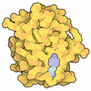
| Title | Monomeric crystal structure of RpBphP1 photosensory core domain from the bacterium Rhodopseudomonas palustris | |||||||||
 Components Components | BphP1 | |||||||||
 Keywords Keywords | PHOTOSYNTHESIS / Bacteriophytochrome / Gene Repressor / Biliverdin | |||||||||
| Function / homology |  Function and homology information Function and homology informationdetection of visible light / phosphorelay signal transduction system / photoreceptor activity / kinase activity / regulation of DNA-templated transcription / ATP binding Similarity search - Function | |||||||||
| Biological species |  Rhodopseudomonas palustris (phototrophic) Rhodopseudomonas palustris (phototrophic) | |||||||||
| Method |  X-RAY DIFFRACTION / X-RAY DIFFRACTION /  SYNCHROTRON / SYNCHROTRON /  MOLECULAR REPLACEMENT / Resolution: 2.6 Å MOLECULAR REPLACEMENT / Resolution: 2.6 Å | |||||||||
 Authors Authors | Papiz, M.Z. / Bellini, D. | |||||||||
| Funding support |  United Kingdom, 2items United Kingdom, 2items
| |||||||||
 Citation Citation |  Journal: To Be Published Journal: To Be PublishedTitle: Insights into the light-induced molecular switch of the bacteriophytochrome RpBphP1 probed by SAXS, modelling and UV-Vis optical properties Authors: Papiz, M.Z. / Bellini, D. #1:  Journal: Structure / Year: 2012 Journal: Structure / Year: 2012Title: Structure of a bacteriophytochrome and light-stimulated protomer swapping with a gene repressor. Authors: Bellini, D. / Papiz, M.Z. | |||||||||
| History |
|
- Structure visualization
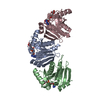
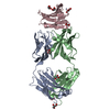
| Structure viewer | Molecule:  Molmil Molmil Jmol/JSmol Jmol/JSmol |
|---|
- Downloads & links
- Download
| PDBx/mmCIF format |  5oy5.cif.gz 5oy5.cif.gz | 216.2 KB | Display |  PDBx/mmCIF format PDBx/mmCIF format |
|---|---|---|---|---|
| PDB format |  pdb5oy5.ent.gz pdb5oy5.ent.gz | 173.3 KB | Display |  PDB format PDB format |
| PDBx/mmJSON format |  5oy5.json.gz 5oy5.json.gz | Tree view |  PDBx/mmJSON format PDBx/mmJSON format | |
| Others |  Other downloads Other downloads |
-Validation report
| Summary document |  5oy5_validation.pdf.gz 5oy5_validation.pdf.gz | 797.3 KB | Display |  wwPDB validaton report wwPDB validaton report |
|---|---|---|---|---|
| Full document |  5oy5_full_validation.pdf.gz 5oy5_full_validation.pdf.gz | 821.4 KB | Display | |
| Data in XML |  5oy5_validation.xml.gz 5oy5_validation.xml.gz | 25.3 KB | Display | |
| Data in CIF |  5oy5_validation.cif.gz 5oy5_validation.cif.gz | 35.5 KB | Display | |
| Arichive directory |  https://data.pdbj.org/pub/pdb/validation_reports/oy/5oy5 https://data.pdbj.org/pub/pdb/validation_reports/oy/5oy5 ftp://data.pdbj.org/pub/pdb/validation_reports/oy/5oy5 ftp://data.pdbj.org/pub/pdb/validation_reports/oy/5oy5 | HTTPS FTP |
-Related structure data
| Related structure data |  4gw9S S: Starting model for refinement |
|---|---|
| Similar structure data |
- Links
Links
- Assembly
| Deposited unit | 
| ||||||||
|---|---|---|---|---|---|---|---|---|---|
| 1 |
| ||||||||
| Unit cell |
|
- Components
| #1: Protein | Mass: 57772.582 Da / Num. of mol.: 1 Source method: isolated from a genetically manipulated source Source: (gene. exp.)  Rhodopseudomonas palustris (strain ATCC BAA-98 / CGA009) (phototrophic) Rhodopseudomonas palustris (strain ATCC BAA-98 / CGA009) (phototrophic)Strain: ATCC BAA-98 / CGA009 / Plasmid: pET28a / Production host:  |
|---|---|
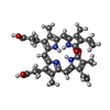
| #2: Chemical | ChemComp-BLA / |
| #3: Water | ChemComp-HOH / |
| Has protein modification | Y |
-Experimental details
-Experiment
| Experiment | Method:  X-RAY DIFFRACTION / Number of used crystals: 1 X-RAY DIFFRACTION / Number of used crystals: 1 |
|---|
- Sample preparation
| Crystal | Density Matthews: 5.07 Å3/Da / Density % sol: 75.77 % / Description: Bipyramid 0.6 mm |
|---|---|
| Crystal grow | Temperature: 292 K / Method: vapor diffusion, hanging drop / pH: 8 Details: 4% PGA (poly-gamma-glutamic acid polymer), 200 mM KBr and 100 mM TrisHCl pH 8 |
-Data collection
| Diffraction | Mean temperature: 100 K |
|---|---|
| Diffraction source | Source:  SYNCHROTRON / Site: SYNCHROTRON / Site:  Diamond Diamond  / Beamline: I02 / Wavelength: 0.9763 Å / Beamline: I02 / Wavelength: 0.9763 Å |
| Detector | Type: DECTRIS PILATUS3 S 6M / Detector: PIXEL / Date: Sep 23, 2010 Details: Kirkpatrick Baez (KB) bimorph mirror pair for horizontal and vertical focussing |
| Radiation | Monochromator: double crystal Si(111) / Protocol: SINGLE WAVELENGTH / Monochromatic (M) / Laue (L): M / Scattering type: x-ray |
| Radiation wavelength | Wavelength: 0.9763 Å / Relative weight: 1 |
| Reflection | Resolution: 2.6→79 Å / Num. obs: 34645 / % possible obs: 99.5 % / Observed criterion σ(F): 0 / Redundancy: 11 % / Rsym value: 0.062 / Net I/σ(I): 8.5 |
| Reflection shell | Resolution: 2.6→2.67 Å / Redundancy: 12 % / Rmerge(I) obs: 0.68 / Mean I/σ(I) obs: 1.7 / Num. unique obs: 2758 / Rsym value: 0.62 / % possible all: 99.4 |
- Processing
| Software |
| ||||||||||||||||||||||||||||||||||||||||||||||||||||||||||||||||||||||||||||||||||||||||||||||||||||||||||||||||||||||||||||||||||||||||||||||||||||||||||||||||||||||||||||||||||||||
|---|---|---|---|---|---|---|---|---|---|---|---|---|---|---|---|---|---|---|---|---|---|---|---|---|---|---|---|---|---|---|---|---|---|---|---|---|---|---|---|---|---|---|---|---|---|---|---|---|---|---|---|---|---|---|---|---|---|---|---|---|---|---|---|---|---|---|---|---|---|---|---|---|---|---|---|---|---|---|---|---|---|---|---|---|---|---|---|---|---|---|---|---|---|---|---|---|---|---|---|---|---|---|---|---|---|---|---|---|---|---|---|---|---|---|---|---|---|---|---|---|---|---|---|---|---|---|---|---|---|---|---|---|---|---|---|---|---|---|---|---|---|---|---|---|---|---|---|---|---|---|---|---|---|---|---|---|---|---|---|---|---|---|---|---|---|---|---|---|---|---|---|---|---|---|---|---|---|---|---|---|---|---|---|
| Refinement | Method to determine structure:  MOLECULAR REPLACEMENT MOLECULAR REPLACEMENTStarting model: 4GW9 Resolution: 2.6→79.09 Å / Cor.coef. Fo:Fc: 0.958 / Cor.coef. Fo:Fc free: 0.932 / SU B: 15.214 / SU ML: 0.174 / Cross valid method: THROUGHOUT / ESU R: 0.238 / ESU R Free: 0.219 / Stereochemistry target values: MAXIMUM LIKELIHOOD / Details: HYDROGENS HAVE BEEN USED IF PRESENT IN THE INPUT
| ||||||||||||||||||||||||||||||||||||||||||||||||||||||||||||||||||||||||||||||||||||||||||||||||||||||||||||||||||||||||||||||||||||||||||||||||||||||||||||||||||||||||||||||||||||||
| Solvent computation | Ion probe radii: 0.8 Å / Shrinkage radii: 0.8 Å / VDW probe radii: 1.2 Å / Solvent model: MASK | ||||||||||||||||||||||||||||||||||||||||||||||||||||||||||||||||||||||||||||||||||||||||||||||||||||||||||||||||||||||||||||||||||||||||||||||||||||||||||||||||||||||||||||||||||||||
| Displacement parameters | Biso mean: 77.791 Å2
| ||||||||||||||||||||||||||||||||||||||||||||||||||||||||||||||||||||||||||||||||||||||||||||||||||||||||||||||||||||||||||||||||||||||||||||||||||||||||||||||||||||||||||||||||||||||
| Refinement step | Cycle: 1 / Resolution: 2.6→79.09 Å
| ||||||||||||||||||||||||||||||||||||||||||||||||||||||||||||||||||||||||||||||||||||||||||||||||||||||||||||||||||||||||||||||||||||||||||||||||||||||||||||||||||||||||||||||||||||||
| Refine LS restraints |
|
 Movie
Movie Controller





 PDBj
PDBj
