Mass: 18.015 Da / Num. of mol.: 782 / Source method: isolated from a natural source / Formula: H2O
-
Experimental details
-
Experiment
Experiment
Method: X-RAY DIFFRACTION / Number of used crystals: 1
-
Sample preparation
Crystal
Density Matthews: 2.76 Å3/Da / Density % sol: 55.52 % / Description: Rice-shaped crystals
Crystal grow
Temperature: 293 K / Method: vapor diffusion, hanging drop / pH: 6.3 Details: 50 mM citrate buffer at pH 6.3, containing 1.6 - 2.0 M ammonium sulfate as a precipitant PH range: 6.3-6.7
-
Data collection
Diffraction
Mean temperature: 100 K
Diffraction source
Source: SYNCHROTRON / Site: PETRA III, EMBL c/o DESY / Beamline: P13 (MX1) / Wavelength: 0.934 Å
Resolution: 1.28→114.09 Å / Cor.coef. Fo:Fc: 0.985 / Cor.coef. Fo:Fc free: 0.981 / SU B: 1.352 / SU ML: 0.024 / Cross valid method: THROUGHOUT / ESU R: 0.032 / ESU R Free: 0.032 / Details: HYDROGENS HAVE BEEN ADDED IN THE RIDING POSITIONS
Rfactor
Num. reflection
% reflection
Selection details
Rfree
0.13684
12246
5 %
RANDOM
Rwork
0.11903
-
-
-
obs
0.11992
231654
99.26 %
-
Solvent computation
Ion probe radii: 0.7 Å / Shrinkage radii: 0.7 Å / VDW probe radii: 1.3 Å
 Movie
Movie Controller

 Yorodumi
 Open data
 Basic information
 Components
 Keywords
 Function and homology information
 Sporosarcina pasteurii (bacteria)
 X-RAY DIFFRACTION /
  Authors
 Citation
 Structure visualization
 Downloads & links
 Other downloads

 PDBj
PDBj

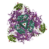
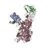
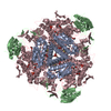
 Assembly
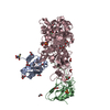


 Mass: 62.068 Da / Num. of mol.: 17 / Source method: obtained synthetically / Formula: C2H6O2
 Mass: 96.063 Da / Num. of mol.: 3 / Source method: obtained synthetically / Formula: SO4
 Mass: 58.693 Da / Num. of mol.: 2 / Source method: obtained synthetically / Formula: Ni
 Mass: 113.076 Da / Num. of mol.: 1 / Source method: isolated from a natural source / Formula: H4NO2PS / Feature type: SUBJECT OF INVESTIGATION
 Sample preparation
 / Beamline: P13 (MX1) / Wavelength: 0.934 Å
 Processing