 Movie
Movie Controller


+ Open data

- Basic information
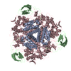
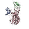
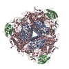
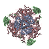
| Entry | Database: PDB / ID: 1a5l | ||||||
|---|---|---|---|---|---|---|---|
| Title | K217C VARIANT OF KLEBSIELLA AEROGENES UREASE | ||||||
 Components Components |
| ||||||
 Keywords Keywords | HYDROLASE / HYDROLASE (UREA AMIDO) / MUTANT / NICKEL METALLOENZYME | ||||||
| Function / homology |  Function and homology information Function and homology informationurease complex / urease / urease activity / urea catabolic process / nickel cation binding / cytoplasm Similarity search - Function | ||||||
| Biological species |  Klebsiella aerogenes (bacteria) Klebsiella aerogenes (bacteria) | ||||||
| Method |  X-RAY DIFFRACTION / DIFFERENCE FOURIER / Resolution: 2.2 Å X-RAY DIFFRACTION / DIFFERENCE FOURIER / Resolution: 2.2 Å | ||||||
 Authors Authors | Pearson, M.A. / Schaller, R.A. / Michel, L.O. / Karplus, P.A. / Hausinger, R.P. | ||||||
 Citation Citation | Journal: Biochemistry / Year: 1998 Title: Chemical rescue of Klebsiella aerogenes urease variants lacking the carbamylated-lysine nickel ligand. Authors: Pearson, M.A. / Schaller, R.A. / Michel, L.O. / Karplus, P.A. / Hausinger, R.P. #1: Journal: Acc.Chem.Res. / Year: 1997Title: 70 Years of Crystalline Urease: What Have We Learned? Authors: Karplus, P.A. / Pearson, M.A. / Hausinger, R.P. #2: Journal: Science / Year: 1995Title: The Crystal Structure of Urease from Klebsiella Aerogenes Authors: Jabri, E. / Carr, M.B. / Hausinger, R.P. / Karplus, P.A. | ||||||
| History |
|
- Structure visualization
| Structure viewer | Molecule: MolmilJmol/JSmol |
|---|
- Downloads & links
-Download
| PDBx/mmCIF format | 1a5l.cif.gz | 151.9 KB | Display | PDBx/mmCIF format |
|---|---|---|---|---|
| PDB format | pdb1a5l.ent.gz | 118.8 KB | Display | PDB format |
| PDBx/mmJSON format | 1a5l.json.gz | Tree view | PDBx/mmJSON format | |
| Others |  Other downloads Other downloads |
-Validation report
| Arichive directory | https://data.pdbj.org/pub/pdb/validation_reports/a5/1a5lftp://data.pdbj.org/pub/pdb/validation_reports/a5/1a5l | HTTPS FTP |
|---|
-Related structure data


-Links
 PDBj
PDBj

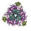
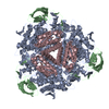
- Assembly
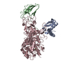
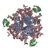
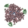
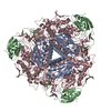
| Deposited unit | 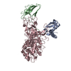
| ||||||||
|---|---|---|---|---|---|---|---|---|---|
| 1 | 
| ||||||||
| Unit cell |
|
-Components
| #1: Protein | Mass: 11100.928 Da / Num. of mol.: 1 / Mutation: K217C, C319A Source method: isolated from a genetically manipulated source Source: (gene. exp.) Klebsiella aerogenes (bacteria) / Gene: UREA, UREB, UREC / Plasmid: PKAU17 / Gene (production host): UREA, UREB, UREC / Production host: |
|---|---|
| #2: Protein | Mass: 11125.690 Da / Num. of mol.: 1 Source method: isolated from a genetically manipulated source Source: (gene. exp.) Klebsiella aerogenes (bacteria) / Gene: UREA, UREB, UREC / Plasmid: PKAU17 / Gene (production host): UREA, UREB, UREC / Production host: |
| #3: Protein | Mass: 60209.125 Da / Num. of mol.: 1 Source method: isolated from a genetically manipulated source Source: (gene. exp.) Klebsiella aerogenes (bacteria) / Gene: UREA, UREB, UREC / Plasmid: PKAU17 / Gene (production host): UREA, UREB, UREC / Production host: |
| #4: Water | ChemComp-HOH /  Mass: 18.015 Da / Num. of mol.: 183 / Source method: isolated from a natural source / Formula: H2O Mass: 18.015 Da / Num. of mol.: 183 / Source method: isolated from a natural source / Formula: H2O |
-Experimental details
-Experiment
| Experiment | Method: X-RAY DIFFRACTION / Number of used crystals: 1 |
|---|
- Sample preparation
| Crystal | Density Matthews: 2.52 Å3/Da / Density % sol: 49 % | ||||||||||||||||||||||||||||||||||||||||||||||||||||||
|---|---|---|---|---|---|---|---|---|---|---|---|---|---|---|---|---|---|---|---|---|---|---|---|---|---|---|---|---|---|---|---|---|---|---|---|---|---|---|---|---|---|---|---|---|---|---|---|---|---|---|---|---|---|---|---|
| Crystal grow | pH: 7.5 Details: PROTEIN WAS CRYSTALLIZED FROM 100 MM HEPES, PH 7.5, 1.6 M LI2SO4 | ||||||||||||||||||||||||||||||||||||||||||||||||||||||
| Crystal grow | *PLUS Temperature: 25 ℃ / Method: vapor diffusion, hanging drop | ||||||||||||||||||||||||||||||||||||||||||||||||||||||
| Components of the solutions | *PLUS
|
-Data collection
| Diffraction | Mean temperature: 298 K |
|---|---|
| Diffraction source | Source: ROTATING ANODE / Type: RIGAKU / Wavelength: 1.5418 |
| Detector | Type: XUONG-HAMLIN MULTIWIRE / Detector: AREA DETECTOR / Date: Oct 1, 1997 |
| Radiation | Monochromatic (M) / Laue (L): M / Scattering type: x-ray |
| Radiation wavelength | Wavelength: 1.5418 Å / Relative weight: 1 |
| Reflection | Highest resolution: 2.2 Å / Num. obs: 38169 / % possible obs: 93 % / Redundancy: 2 % / Rsym value: 0.091 / Net I/σ(I): 7.7 |
| Reflection shell | Resolution: 2.2→2.28 Å / Redundancy: 1.3 % / Mean I/σ(I) obs: 2.6 / Rsym value: 0.288 / % possible all: 69 |
| Reflection | *PLUS Rmerge(I) obs: 0.091 |
| Reflection shell | *PLUS % possible obs: 69 % / Rmerge(I) obs: 0.288 |
- Processing
| Software |
| ||||||||||||||||||||||||||||||||||||||||||||||||||||||||||||
|---|---|---|---|---|---|---|---|---|---|---|---|---|---|---|---|---|---|---|---|---|---|---|---|---|---|---|---|---|---|---|---|---|---|---|---|---|---|---|---|---|---|---|---|---|---|---|---|---|---|---|---|---|---|---|---|---|---|---|---|---|---|
| Refinement | Method to determine structure: DIFFERENCE FOURIER / Resolution: 2.2→10 Å / σ(F): 0 Details: ALL NON-BONDED INTERACTIONS WERE REMOVED BETWEEN THE TWO ALTERNATE CONFORMATIONS OF HIS272 OF CHAIN C THE OCCUPANCIES FOR THE TWO ALTERNATE CONFORMATIONS OBSERVED FOR HIS 272 WERE REFINED ...Details: ALL NON-BONDED INTERACTIONS WERE REMOVED BETWEEN THE TWO ALTERNATE CONFORMATIONS OF HIS272 OF CHAIN C THE OCCUPANCIES FOR THE TWO ALTERNATE CONFORMATIONS OBSERVED FOR HIS 272 WERE REFINED WITH A FIXED B-FACTOR OF 15 (ANGSTROMS)**2.
| ||||||||||||||||||||||||||||||||||||||||||||||||||||||||||||
| Refinement step | Cycle: LAST / Resolution: 2.2→10 Å
| ||||||||||||||||||||||||||||||||||||||||||||||||||||||||||||
| Refine LS restraints |
| ||||||||||||||||||||||||||||||||||||||||||||||||||||||||||||
| LS refinement shell | Resolution: 2.2→2.3 Å / Total num. of bins used: 10
| ||||||||||||||||||||||||||||||||||||||||||||||||||||||||||||
| Xplor file |
| ||||||||||||||||||||||||||||||||||||||||||||||||||||||||||||
| Software | *PLUS Name: X-PLOR / Version: 3.1 / Classification: refinement | ||||||||||||||||||||||||||||||||||||||||||||||||||||||||||||
| Refinement | *PLUS | ||||||||||||||||||||||||||||||||||||||||||||||||||||||||||||
| Solvent computation | *PLUS | ||||||||||||||||||||||||||||||||||||||||||||||||||||||||||||
| Displacement parameters | *PLUS | ||||||||||||||||||||||||||||||||||||||||||||||||||||||||||||
| Refine LS restraints | *PLUS
| ||||||||||||||||||||||||||||||||||||||||||||||||||||||||||||
| LS refinement shell | *PLUS Rfactor obs: 0.259 |
