 Movie
Movie Controller


+ Open data

- Basic information
| Entry | Database: PDB / ID: 1fwi | ||||||
|---|---|---|---|---|---|---|---|
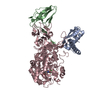
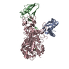
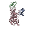
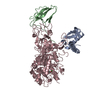
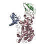
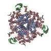
| Title | KLEBSIELLA AEROGENES UREASE, H134A VARIANT | ||||||
 Components Components | (UREASE) x 3 | ||||||
 Keywords Keywords | HYDROLASE / HYDROLASE(UREA AMIDO) / MUTANT / NICKEL METALLOENZYME | ||||||
| Function / homology |  Function and homology information Function and homology informationurease complex / urease / urease activity / urea catabolic process / nickel cation binding / cytoplasm Similarity search - Function | ||||||
| Biological species |  Klebsiella aerogenes (bacteria) Klebsiella aerogenes (bacteria) | ||||||
| Method |  X-RAY DIFFRACTION / Resolution: 2 Å X-RAY DIFFRACTION / Resolution: 2 Å | ||||||
 Authors Authors | Pearson, M.A. / Karplus, P.A. | ||||||
 Citation Citation | Journal: J.Biol.Chem. / Year: 1996 Title: Characterization of the mononickel metallocenter in H134A mutant urease. Authors: Park, I.S. / Michel, L.O. / Pearson, M.A. / Jabri, E. / Karplus, P.A. / Wang, S. / Dong, J. / Scott, R.A. / Koehler, B.P. / Johnson, M.K. / Hausinger, R.P. #1: Journal: Biochemistry / Year: 1996Title: Structures of the Klebsiella Aerogenes Urease Apoenzyme and Two Active-Site Mutants Authors: Jabri, E. / Karplus, P.A. #2: Journal: Science / Year: 1995Title: The Crystal Structure of Urease from Klebsiella Aerogenes Authors: Jabri, E. / Carr, M.B. / Hausinger, R.P. / Karplus, P.A. #3: Journal: Protein Sci. / Year: 1993Title: Site-Directed Mutagenesis of Klebsiella Aerogenes Urease: Identification of Histidine Residues that Appear to Function in Nickel Ligation, Substrate Binding, and Catalysis Authors: Park, I.S. / Hausinger, R.P. | ||||||
| History |
|
- Structure visualization
| Structure viewer | Molecule: MolmilJmol/JSmol |
|---|
- Downloads & links
-Download
| PDBx/mmCIF format | 1fwi.cif.gz | 155.8 KB | Display | PDBx/mmCIF format |
|---|---|---|---|---|
| PDB format | pdb1fwi.ent.gz | 126.5 KB | Display | PDB format |
| PDBx/mmJSON format | 1fwi.json.gz | Tree view | PDBx/mmJSON format | |
| Others |  Other downloads Other downloads |
-Validation report
| Arichive directory | https://data.pdbj.org/pub/pdb/validation_reports/fw/1fwiftp://data.pdbj.org/pub/pdb/validation_reports/fw/1fwi | HTTPS FTP |
|---|
-Related structure data
| Similar structure data |
|---|

-Links
 PDBj
PDBj

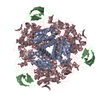
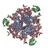
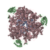
- Assembly
| Deposited unit | 
| ||||||||
|---|---|---|---|---|---|---|---|---|---|
| 1 | 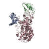
| ||||||||
| Unit cell |
|
-Components
| #1: Protein | Mass: 11100.928 Da / Num. of mol.: 1 / Mutation: H(C 134)A Source method: isolated from a genetically manipulated source Source: (gene. exp.) Klebsiella aerogenes (bacteria) / Plasmid: PKAU17 / Production host: |
|---|---|
| #2: Protein | Mass: 11712.239 Da / Num. of mol.: 1 / Mutation: H(C 134)A Source method: isolated from a genetically manipulated source Source: (gene. exp.) Klebsiella aerogenes (bacteria) / Plasmid: PKAU17 / Production host: |
| #3: Protein | Mass: 60342.289 Da / Num. of mol.: 1 / Mutation: H(C 134)A Source method: isolated from a genetically manipulated source Source: (gene. exp.) Klebsiella aerogenes (bacteria) / Plasmid: PKAU17 / Production host: |
| #4: Chemical | ChemComp-NI /   Mass: 58.693 Da / Num. of mol.: 1 / Source method: obtained synthetically / Formula: Ni Mass: 58.693 Da / Num. of mol.: 1 / Source method: obtained synthetically / Formula: Ni |
| #5: Water | ChemComp-HOH /  Mass: 18.015 Da / Num. of mol.: 308 / Source method: isolated from a natural source / Formula: H2O Mass: 18.015 Da / Num. of mol.: 308 / Source method: isolated from a natural source / Formula: H2O |
| Compound details | THIS MODEL IS THAT OF THE H134A MUTANT AT 2.0 ANGSTROMS. THREE NONIDENTICAL CHAINS, GAMMA (A), BETA ...THIS MODEL IS THAT OF THE H134A MUTANT AT 2.0 ANGSTROMS. THREE NONIDENTIC |
-Experimental details
-Experiment
| Experiment | Method: X-RAY DIFFRACTION |
|---|
- Sample preparation
| Crystal | Density Matthews: 2.5 Å3/Da / Density % sol: 49 % | ||||||||||||||||||||||||||||||||||||||||||||||||||||||
|---|---|---|---|---|---|---|---|---|---|---|---|---|---|---|---|---|---|---|---|---|---|---|---|---|---|---|---|---|---|---|---|---|---|---|---|---|---|---|---|---|---|---|---|---|---|---|---|---|---|---|---|---|---|---|---|
| Crystal grow | *PLUS Temperature: 25 ℃ / pH: 7 / Method: vapor diffusion, hanging drop / Details: Jabri, E., (1992) J.Mol.Biol., 227, 934. | ||||||||||||||||||||||||||||||||||||||||||||||||||||||
| Components of the solutions | *PLUS
|
-Data collection
| Diffraction source | Wavelength: 1.5418 |
|---|---|
| Detector | Type: XUONG-HAMLIN MULTIWIRE / Detector: AREA DETECTOR |
| Radiation | Monochromatic (M) / Laue (L): M / Scattering type: x-ray |
| Radiation wavelength | Wavelength: 1.5418 Å / Relative weight: 1 |
| Reflection | Num. obs: 52421 / % possible obs: 96 % / Observed criterion σ(I): 0 / Redundancy: 3.3 % / Rmerge(I) obs: 0.066 |
| Reflection | *PLUS Highest resolution: 2 Å |
- Processing
| Software |
| ||||||||||||||||||||||||||||||||||||||||||||||||||||||||||||
|---|---|---|---|---|---|---|---|---|---|---|---|---|---|---|---|---|---|---|---|---|---|---|---|---|---|---|---|---|---|---|---|---|---|---|---|---|---|---|---|---|---|---|---|---|---|---|---|---|---|---|---|---|---|---|---|---|---|---|---|---|---|
| Refinement | Resolution: 2→10 Å / σ(F): 0
| ||||||||||||||||||||||||||||||||||||||||||||||||||||||||||||
| Refinement step | Cycle: LAST / Resolution: 2→10 Å
| ||||||||||||||||||||||||||||||||||||||||||||||||||||||||||||
| Refine LS restraints |
| ||||||||||||||||||||||||||||||||||||||||||||||||||||||||||||
| Software | *PLUS Name: X-PLOR / Version: 3.1 / Classification: refinement | ||||||||||||||||||||||||||||||||||||||||||||||||||||||||||||
| Refinement | *PLUS Rfactor obs: 0.17 / Rfactor Rwork: 0.17 | ||||||||||||||||||||||||||||||||||||||||||||||||||||||||||||
| Solvent computation | *PLUS | ||||||||||||||||||||||||||||||||||||||||||||||||||||||||||||
| Displacement parameters | *PLUS |
