 Movie
Movie Controller


+ Open data

- Basic information
| Entry | Database: PDB / ID: 1ie7 | ||||||
|---|---|---|---|---|---|---|---|
| Title | PHOSPHATE INHIBITED BACILLUS PASTEURII UREASE CRYSTAL STRUCTURE | ||||||
 Components Components |
| ||||||
 Keywords Keywords | HYDROLASE / UREASE / BACILLUS PASTEURII / NICKEL / METALLOENZYME | ||||||
| Function / homology |  Function and homology information Function and homology informationurease complex / urease / urease activity / urea catabolic process / nickel cation binding / cytoplasm Similarity search - Function | ||||||
| Biological species |  Sporosarcina pasteurii (bacteria) Sporosarcina pasteurii (bacteria) | ||||||
| Method |  X-RAY DIFFRACTION / SYNCHROTRON / MOLECULAR REPLACEMENT / Resolution: 1.85 Å X-RAY DIFFRACTION / SYNCHROTRON / MOLECULAR REPLACEMENT / Resolution: 1.85 Å | ||||||
 Authors Authors | Benini, S. / Rypniewski, W.R. / Wilson, K.S. / Ciurli, S. / Mangani, S. | ||||||
 Citation Citation | Journal: J.Biol.Inorg.Chem. / Year: 2001 Title: Structure-based rationalization of urease inhibition by phosphate: novel insights into the enzyme mechanism. Authors: Benini, S. / Rypniewski, W.R. / Wilson, K.S. / Ciurli, S. / Mangani, S. #1: Journal: J.BIOL.INORG.CHEM. / Year: 2000Title: The Complex of Bacillus pasteurii Urease with Acetohydroxamate anion from X-ray Data at 1.55 A Resolution Authors: Benini, S. / Rypniewski, W.R. / Wilson, K.S. / Miletti, S. / Ciurli, S. / Mangani, S. #2: Journal: Structure / Year: 1999Title: A New Proposal for Urease Mechanism Based on the Crystal Structures of the Native and Inhibited Enzyme from Bacillus pasteurii: Why Urea Hydrolysis Costs Two Nickels Authors: Benini, S. / Rypniewski, W.R. / Wilson, K.S. / Miletti, S. / Ciurli, S. / Mangani, S. #3: Journal: J.BIOL.INORG.CHEM. / Year: 1998Title: The Complex of Bacillus pasteurii Urease with beta-mercaptoethanol from X-ray Data at 1.65 A Resolution Authors: Benini, S. / Ciurli, S. / Rypniewski, W.R. / Wilson, K.S. / Mangani, S. #4: Journal: Acta Crystallogr.,Sect.D / Year: 1998Title: Crystallization and Preliminary High-resolution X-ray Diffraction Analysis of Native and beta-mercaptoethanol-inhibited Urease from Bacillus pasteurii Authors: Benini, S. / Ciurli, S. / Rypniewski, W.R. / Wilson, K.S. / Mangani, S. | ||||||
| History |
| ||||||
| Remark 999 | SEQUENCE There are differences between the original sequence from DNA reported by others and the ...SEQUENCE There are differences between the original sequence from DNA reported by others and the electron density maps, including an insertion. The modified residues are due to posttranslational modifications. |
- Structure visualization
| Structure viewer | Molecule: MolmilJmol/JSmol |
|---|
- Downloads & links
-Download
| PDBx/mmCIF format | 1ie7.cif.gz | 191 KB | Display | PDBx/mmCIF format |
|---|---|---|---|---|
| PDB format | pdb1ie7.ent.gz | 146.1 KB | Display | PDB format |
| PDBx/mmJSON format | 1ie7.json.gz | Tree view | PDBx/mmJSON format | |
| Others |  Other downloads Other downloads |
-Validation report
| Arichive directory | https://data.pdbj.org/pub/pdb/validation_reports/ie/1ie7ftp://data.pdbj.org/pub/pdb/validation_reports/ie/1ie7 | HTTPS FTP |
|---|
-Related structure data
| Related structure data |  2ubpS S: Starting model for refinement |
|---|---|
| Similar structure data |


-Links
 PDBj
PDBj

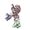
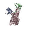
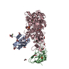
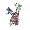
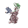
- Assembly
| Deposited unit | 
| ||||||||
|---|---|---|---|---|---|---|---|---|---|
| 1 |
| ||||||||
| 2 | 
| ||||||||
| 3 | 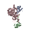
| ||||||||
| Unit cell |
|
-Components
-Protein , 3 types, 3 molecules ABC
| #1: Protein | Mass: 11204.988 Da / Num. of mol.: 1 / Source method: isolated from a natural source / Source: (natural) Sporosarcina pasteurii (bacteria) / Strain: DSM 33 / References: UniProt: P41022, urease |
|---|---|
| #2: Protein | Mass: 13975.539 Da / Num. of mol.: 1 / Source method: isolated from a natural source / Source: (natural) Sporosarcina pasteurii (bacteria) / Strain: DSM 33 / References: UniProt: P41021, urease |
| #3: Protein | Mass: 61632.742 Da / Num. of mol.: 1 / Source method: isolated from a natural source / Source: (natural) Sporosarcina pasteurii (bacteria) / Strain: DSM 33 / References: UniProt: P41020, urease |
-Non-polymers , 3 types, 899 molecules 


| #4: Chemical |  Mass: 58.693 Da / Num. of mol.: 2 / Source method: obtained synthetically / Formula: Ni Mass: 58.693 Da / Num. of mol.: 2 / Source method: obtained synthetically / Formula: Ni#5: Chemical | ChemComp-PO4 / |  Mass: 94.971 Da / Num. of mol.: 1 / Source method: obtained synthetically / Formula: PO4 Mass: 94.971 Da / Num. of mol.: 1 / Source method: obtained synthetically / Formula: PO4#6: Water | ChemComp-HOH / | Mass: 18.015 Da / Num. of mol.: 896 / Source method: isolated from a natural source / Formula: H2O |
|---|
-Experimental details
-Experiment
| Experiment | Method: X-RAY DIFFRACTION / Number of used crystals: 1 |
|---|
- Sample preparation
| Crystal | Density Matthews: 2.99 Å3/Da / Density % sol: 58.58 % | ||||||||||||||||||||||||||||||||||||||||||
|---|---|---|---|---|---|---|---|---|---|---|---|---|---|---|---|---|---|---|---|---|---|---|---|---|---|---|---|---|---|---|---|---|---|---|---|---|---|---|---|---|---|---|---|
| Crystal grow | Temperature: 293 K / Method: vapor diffusion, hanging drop / pH: 6.3 Details: 1.8 M AMS, 100 mM Sodium Phosphate, pH 6.3, VAPOR DIFFUSION, HANGING DROP, temperature 293K | ||||||||||||||||||||||||||||||||||||||||||
| Crystal grow | *PLUS Temperature: 20 K / pH: 6.8 | ||||||||||||||||||||||||||||||||||||||||||
| Components of the solutions | *PLUS
|
-Data collection
| Diffraction | Mean temperature: 100 K |
|---|---|
| Diffraction source | Source: SYNCHROTRON / Site: EMBL/DESY, HAMBURG  / Beamline: BW7B / Wavelength: 0.834 Å / Beamline: BW7B / Wavelength: 0.834 Å |
| Detector | Type: MARRESEARCH / Detector: IMAGE PLATE / Date: Jun 10, 1998 / Details: BENT MIRROR |
| Radiation | Monochromator: SI(111) / Protocol: SINGLE WAVELENGTH / Monochromatic (M) / Laue (L): M / Scattering type: x-ray |
| Radiation wavelength | Wavelength: 0.834 Å / Relative weight: 1 |
| Reflection | Resolution: 1.85→11.987 Å / Num. all: 82718 / Num. obs: 379334 / % possible obs: 99.3 % / Observed criterion σ(F): 2 / Observed criterion σ(I): 2 / Redundancy: 4.58 % / Biso Wilson estimate: 22.08 Å2 / Rmerge(I) obs: 0.097 / Rsym value: 0.097 / Net I/σ(I): 10.2 |
| Reflection shell | Resolution: 1.85→1.88 Å / Redundancy: 3.14 % / Rmerge(I) obs: 0.448 / Mean I/σ(I) obs: 1.85 / Num. unique all: 3824 / Rsym value: 0.448 / % possible all: 94.3 |
| Reflection | *PLUS Lowest resolution: 30 Å / Num. obs: 82718 / Redundancy: 4.61 % / Num. measured all: 381291 |
| Reflection shell | *PLUS % possible obs: 94.3 % / Redundancy: 3.2 % |
- Processing
| Software |
| ||||||||||||||||||||||||||||||||||||||||||||||||
|---|---|---|---|---|---|---|---|---|---|---|---|---|---|---|---|---|---|---|---|---|---|---|---|---|---|---|---|---|---|---|---|---|---|---|---|---|---|---|---|---|---|---|---|---|---|---|---|---|---|
| Refinement | Method to determine structure: MOLECULAR REPLACEMENT Starting model: PDB ENTRY 2ubp Resolution: 1.85→11.99 Å / SU B: 2.8591 / SU ML: 0.0826 / Isotropic thermal model: Isotropic / Cross valid method: THROUGHOUT / σ(F): 2 / σ(I): 2 / ESU R: 0.1183 / ESU R Free: 0.1161 / Stereochemistry target values: Engh & Huber Details: THE PROTEIN ATOMS INVOLVED IN CLOSE CONTACTS WITH WATERS IN REMARK 500 ARE DISORDERED AS INDICATED BY THEIR OCCUPANCY = 0. The residue ASN C 327 that has a chirality error at the CA center ...Details: THE PROTEIN ATOMS INVOLVED IN CLOSE CONTACTS WITH WATERS IN REMARK 500 ARE DISORDERED AS INDICATED BY THEIR OCCUPANCY = 0. The residue ASN C 327 that has a chirality error at the CA center is located in a very disordered region in the electron desity map.
| ||||||||||||||||||||||||||||||||||||||||||||||||
| Displacement parameters | Biso mean: 30.7 Å2 | ||||||||||||||||||||||||||||||||||||||||||||||||
| Refinement step | Cycle: LAST / Resolution: 1.85→11.99 Å
| ||||||||||||||||||||||||||||||||||||||||||||||||
| Refine LS restraints |
| ||||||||||||||||||||||||||||||||||||||||||||||||
| LS refinement shell | Resolution: 1.85→1.93 Å
| ||||||||||||||||||||||||||||||||||||||||||||||||
| Software | *PLUS Name: REFMAC / Classification: refinement | ||||||||||||||||||||||||||||||||||||||||||||||||
| Refinement | *PLUS Lowest resolution: 12 Å / σ(F): 2 / % reflection Rfree: 2 % / Rfactor obs: 0.173 / Rfactor Rfree: 0.21 / Rfactor Rwork: 0.17 | ||||||||||||||||||||||||||||||||||||||||||||||||
| Solvent computation | *PLUS | ||||||||||||||||||||||||||||||||||||||||||||||||
| Displacement parameters | *PLUS Biso mean: 30.7 Å2 | ||||||||||||||||||||||||||||||||||||||||||||||||
| Refine LS restraints | *PLUS
|
