 Movie
Movie Controller

[English] 日本語
 Yorodumi
- PDB-4ac7: The crystal structure of Sporosarcina pasteurii urease in complex... -
+ Open data

- Basic information
| Entry | Database: PDB / ID: 4ac7 | ||||||
|---|---|---|---|---|---|---|---|
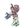
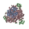
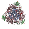
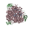
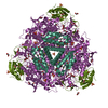
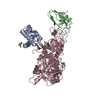
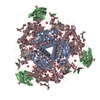
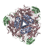
| Title | The crystal structure of Sporosarcina pasteurii urease in complex with citrate | ||||||
 Components Components | (UREASE SUBUNIT ...) x 3 | ||||||
 Keywords Keywords | HYDROLASE / BACILLUS PASTEURII | ||||||
| Function / homology |  Function and homology information Function and homology informationurease complex / urease / urease activity / urea catabolic process / nickel cation binding / cytoplasm Similarity search - Function | ||||||
| Biological species |  SPOROSARCINA PASTEURII (bacteria) SPOROSARCINA PASTEURII (bacteria) | ||||||
| Method |  X-RAY DIFFRACTION / SYNCHROTRON / FOURIER SYNTHESIS / Resolution: 1.5 Å X-RAY DIFFRACTION / SYNCHROTRON / FOURIER SYNTHESIS / Resolution: 1.5 Å | ||||||
 Authors Authors | Benini, S. / Kosikowska, P. / Cianci, M. / Gonzalez Vara, A. / Berlicki, L. / Ciurli, S. | ||||||
 Citation Citation | Journal: J.Biol.Inorg.Chem. / Year: 2013 Title: The Crystal Structure of Sporosarcina Pasteurii Urease in a Complex with Citrate Provides New Hints for Inhibitor Design. Authors: Benini, S. / Kosikowska, P. / Cianci, M. / Mazzei, L. / Vara, A.G. / Berlicki, L. / Ciurli, S. #1: Journal: J.Am.Chem.Soc. / Year: 2004Title: Molecular Details of Urease Inhibition by Boric Acid: Insights Into the Catalytic Mechanism. Authors: Benini, S. / Rypniewski, W.R. / Wilson, K.S. / Mangani, S. / Ciurli, S. #2: Journal: J.Biol.Inorg.Chem. / Year: 2001Title: Structure-Based Rationalization of Urease Inhibition by Phosphate: Novel Insights Into the Enzyme Mechanism. Authors: Benini, S. / Rypniewski, W.R. / Wilson, K.S. / Ciurli, S. / Mangani, S. #3: Journal: J.Biol.Inorg.Chem. / Year: 2000Title: The Complex of Bacillus Pasteurii Urease with Acetohydroxamate Anion from X-Ray Data at 1.55 A Resolution. Authors: Benini, S. / Rypniewski, W.R. / Wilson, K.S. / Miletti, S. / Ciurli, S. / Mangani, S. #4: Journal: Structure / Year: 1999Title: A New Proposal for Urease Mechanism Based on the Crystal Structures of the Native and Inhibited Enzyme from Bacillus Pasteurii: Why Urea Hydrolysis Costs Two Nickels. Authors: Benini, S. / Rypniewski, W.R. / Wilson, K.S. / Miletti, S. / Ciurli, S. / Mangani, S. #5: Journal: J.Biol.Inorg.Chem. / Year: 1998Title: The Complex of Bacillus Pasteurii Urease with Beta-Mercaptoethanol from X-Ray Data at 1.65 A Resolution Authors: Benini, S. / Ciurli, S. / Rypniewski, W.R. / Wilson, K.S. / Mangani, S. #6: Journal: Acta Crystallogr.,Sect.D / Year: 1998 Title: Crystallization and Preliminary High-Resolution X-Ray Diffraction Analysis of Native and Beta-Mercaptoethanol-Inhibited Urease from Bacillus Pasteurii. Authors: Benini, S. / Ciurli, S. / Rypniewski, W.R. / Wilson, K.S. / Mangani, S. | ||||||
| History |
|
- Structure visualization
| Structure viewer | Molecule: MolmilJmol/JSmol |
|---|
- Downloads & links
-Download
| PDBx/mmCIF format | 4ac7.cif.gz | 341 KB | Display | PDBx/mmCIF format |
|---|---|---|---|---|
| PDB format | pdb4ac7.ent.gz | 275.3 KB | Display | PDB format |
| PDBx/mmJSON format | 4ac7.json.gz | Tree view | PDBx/mmJSON format | |
| Others |  Other downloads Other downloads |
-Validation report
| Arichive directory | https://data.pdbj.org/pub/pdb/validation_reports/ac/4ac7ftp://data.pdbj.org/pub/pdb/validation_reports/ac/4ac7 | HTTPS FTP |
|---|
-Related structure data
| Related structure data |  1s3tS S: Starting model for refinement |
|---|---|
| Similar structure data |


-Links
 PDBj
PDBj


- Assembly
| Deposited unit | 
| |||||||||
|---|---|---|---|---|---|---|---|---|---|---|
| 1 | 
| |||||||||
| Unit cell |
| |||||||||
| Components on special symmetry positions |
|
-Components
-UREASE SUBUNIT ... , 3 types, 3 molecules ABC
| #1: Protein | Mass: 11204.988 Da / Num. of mol.: 1 / Source method: isolated from a natural source / Source: (natural) SPOROSARCINA PASTEURII (bacteria) / Strain: DSM33 / References: UniProt: P41022, urease |
|---|---|
| #2: Protein | Mass: 13975.539 Da / Num. of mol.: 1 / Source method: isolated from a natural source / Source: (natural) SPOROSARCINA PASTEURII (bacteria) / Strain: DSM33 / References: UniProt: P41021, urease |
| #3: Protein | Mass: 61646.766 Da / Num. of mol.: 1 / Source method: isolated from a natural source / Source: (natural) SPOROSARCINA PASTEURII (bacteria) / Strain: DSM33 / References: UniProt: P41020, urease |
-Non-polymers , 6 types, 689 molecules 


| #4: Chemical | ChemComp-EDO /  Mass: 62.068 Da / Num. of mol.: 10 / Source method: obtained synthetically / Formula: C2H6O2 Mass: 62.068 Da / Num. of mol.: 10 / Source method: obtained synthetically / Formula: C2H6O2#5: Chemical | ChemComp-SO4 /  Mass: 96.063 Da / Num. of mol.: 4 / Source method: obtained synthetically / Formula: SO4 Mass: 96.063 Da / Num. of mol.: 4 / Source method: obtained synthetically / Formula: SO4#6: Chemical |  Mass: 58.693 Da / Num. of mol.: 2 / Source method: obtained synthetically / Formula: Ni Mass: 58.693 Da / Num. of mol.: 2 / Source method: obtained synthetically / Formula: Ni#7: Chemical | ChemComp-OH / |  Mass: 17.007 Da / Num. of mol.: 1 / Source method: obtained synthetically / Formula: HO Mass: 17.007 Da / Num. of mol.: 1 / Source method: obtained synthetically / Formula: HO#8: Chemical | ChemComp-FLC / |  Mass: 189.100 Da / Num. of mol.: 1 / Source method: obtained synthetically / Formula: C6H5O7 Mass: 189.100 Da / Num. of mol.: 1 / Source method: obtained synthetically / Formula: C6H5O7#9: Water | ChemComp-HOH / | Mass: 18.015 Da / Num. of mol.: 671 / Source method: isolated from a natural source / Formula: H2O |
|---|
-Details
| Nonpolymer details | ETHYLENE GLYCOL (EDO): PRESENT AS CRYOPROTECTANT CITRATE ANION (FLC): PRESENT AS A CRYSTALLISATION ...ETHYLENE GLYCOL (EDO): PRESENT AS CRYOPROTEC |
|---|---|
| Sequence details | THE AUTHOR STATES THAT THE RESIDUES LISTED IN SEQADV ARE WHAT IS SEEN IN THE DENSITY, AND IT IS NOT ...THE AUTHOR STATES THAT THE RESIDUES LISTED IN SEQADV ARE WHAT IS SEEN IN THE DENSITY, AND IT IS NOT CLEAR WHERE THE DISCREPANC |
-Experimental details
-Experiment
| Experiment | Method: X-RAY DIFFRACTION / Number of used crystals: 1 |
|---|
- Sample preparation
| Crystal | Density Matthews: 2.76 Å3/Da / Density % sol: 55.46 % |
|---|---|
| Crystal grow | pH: 6.3 Details: 1.8 M AMMONIUM SULPHATE, 100 MM CITRATE PH 6.3, 50 MM NA2SO3 |
-Data collection
| Diffraction | Mean temperature: 100 K |
|---|---|
| Diffraction source | Source: SYNCHROTRON / Site: PETRA III, EMBL c/o DESY  / Beamline: P14 (MX2) / Wavelength: 1.1267 / Beamline: P14 (MX2) / Wavelength: 1.1267 |
| Detector | Type: MARRESEARCH / Detector: CCD / Date: Sep 30, 2011 |
| Radiation | Monochromator: SI(III) CRYSTAL MONOCHROMATOR / Protocol: SINGLE WAVELENGTH / Monochromatic (M) / Laue (L): M / Scattering type: x-ray |
| Radiation wavelength | Wavelength: 1.1267 Å / Relative weight: 1 |
| Reflection | Resolution: 1.5→113.93 Å / Num. obs: 153738 / % possible obs: 99.9 % / Observed criterion σ(I): 2 / Redundancy: 15.5 % / Rmerge(I) obs: 0.1 / Net I/σ(I): 22.6 |
| Reflection shell | Resolution: 1.5→1.58 Å / Redundancy: 9.9 % / Rmerge(I) obs: 0.58 / Mean I/σ(I) obs: 4 / % possible all: 100 |
- Processing
| Software |
| ||||||||||||||||||||||||||||||||||||||||||||||||||||||||||||||||||||||||||||||||||||||||||||||||||||||||||||||||||||||||||||||||||||||||||||||||||||||||||||||||||||||||||||||||||||||
|---|---|---|---|---|---|---|---|---|---|---|---|---|---|---|---|---|---|---|---|---|---|---|---|---|---|---|---|---|---|---|---|---|---|---|---|---|---|---|---|---|---|---|---|---|---|---|---|---|---|---|---|---|---|---|---|---|---|---|---|---|---|---|---|---|---|---|---|---|---|---|---|---|---|---|---|---|---|---|---|---|---|---|---|---|---|---|---|---|---|---|---|---|---|---|---|---|---|---|---|---|---|---|---|---|---|---|---|---|---|---|---|---|---|---|---|---|---|---|---|---|---|---|---|---|---|---|---|---|---|---|---|---|---|---|---|---|---|---|---|---|---|---|---|---|---|---|---|---|---|---|---|---|---|---|---|---|---|---|---|---|---|---|---|---|---|---|---|---|---|---|---|---|---|---|---|---|---|---|---|---|---|---|---|
| Refinement | Method to determine structure: FOURIER SYNTHESIS Starting model: PDB ENTRY 1S3T DEPLETED OF WATERS SULPHATES AND BORATE WAS USED AS A MODEL FOR RIGID BODY REFINEMENT FOLLOWED BY REFINEMENT AND MODEL BUILDING Resolution: 1.5→113.93 Å / Cor.coef. Fo:Fc: 0.981 / Cor.coef. Fo:Fc free: 0.974 / SU B: 1.71 / SU ML: 0.029 / Cross valid method: THROUGHOUT / ESU R: 0.051 / ESU R Free: 0.048 / Stereochemistry target values: MAXIMUM LIKELIHOOD Details: HYDROGENS HAVE BEEN ADDED IN THE RIDING POSITIONS. U VALUES REFINED INDIVIDUALLY
| ||||||||||||||||||||||||||||||||||||||||||||||||||||||||||||||||||||||||||||||||||||||||||||||||||||||||||||||||||||||||||||||||||||||||||||||||||||||||||||||||||||||||||||||||||||||
| Solvent computation | Ion probe radii: 0.8 Å / Shrinkage radii: 0.8 Å / VDW probe radii: 1.2 Å / Solvent model: MASK | ||||||||||||||||||||||||||||||||||||||||||||||||||||||||||||||||||||||||||||||||||||||||||||||||||||||||||||||||||||||||||||||||||||||||||||||||||||||||||||||||||||||||||||||||||||||
| Displacement parameters | Biso mean: 13.109 Å2
| ||||||||||||||||||||||||||||||||||||||||||||||||||||||||||||||||||||||||||||||||||||||||||||||||||||||||||||||||||||||||||||||||||||||||||||||||||||||||||||||||||||||||||||||||||||||
| Refinement step | Cycle: LAST / Resolution: 1.5→113.93 Å
| ||||||||||||||||||||||||||||||||||||||||||||||||||||||||||||||||||||||||||||||||||||||||||||||||||||||||||||||||||||||||||||||||||||||||||||||||||||||||||||||||||||||||||||||||||||||
| Refine LS restraints |
|