 Movie
Movie Controller

[English] 日本語
 Yorodumi
- PDB-5obv: Mycoplasma genitalium DnaK deletion mutant lacking SBDalpha in co... -
+ Open data

- Basic information
| Entry | Database: PDB / ID: 5obv | ||||||
|---|---|---|---|---|---|---|---|
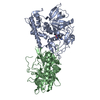
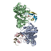
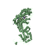
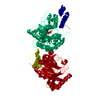
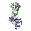
| Title | Mycoplasma genitalium DnaK deletion mutant lacking SBDalpha in complex with ADP and Pi. | ||||||
 Components Components | Chaperone protein DnaK | ||||||
 Keywords Keywords | CHAPERONE / Complex / co-factor / ATP hydrolysis | ||||||
| Function / homology |  Function and homology information Function and homology informationheat shock protein binding / protein folding chaperone / ATP-dependent protein folding chaperone / : / protein refolding / ATP hydrolysis activity / ATP binding Similarity search - Function | ||||||
| Biological species |  Mycoplasma genitalium (bacteria) Mycoplasma genitalium (bacteria) | ||||||
| Method |  X-RAY DIFFRACTION / SYNCHROTRON / MOLECULAR REPLACEMENT / Resolution: 2.49 Å X-RAY DIFFRACTION / SYNCHROTRON / MOLECULAR REPLACEMENT / Resolution: 2.49 Å | ||||||
 Authors Authors | Adell, M. / Calisto, B. / Fita, I. / Martinelli, L. | ||||||
| Funding support |  Spain, 1items Spain, 1items
| ||||||
 Citation Citation | Journal: Protein Sci. / Year: 2018 Title: The nucleotide-bound/substrate-bound conformation of the Mycoplasma genitalium DnaK chaperone. Authors: Adell, M. / Calisto, B.M. / Fita, I. / Martinelli, L. | ||||||
| History |
|
- Structure visualization
| Structure viewer | Molecule: MolmilJmol/JSmol |
|---|
- Downloads & links
-Download
| PDBx/mmCIF format | 5obv.cif.gz | 163.9 KB | Display | PDBx/mmCIF format |
|---|---|---|---|---|
| PDB format | pdb5obv.ent.gz | 123.7 KB | Display | PDB format |
| PDBx/mmJSON format | 5obv.json.gz | Tree view | PDBx/mmJSON format | |
| Others |  Other downloads Other downloads |
-Validation report
| Arichive directory | https://data.pdbj.org/pub/pdb/validation_reports/ob/5obvftp://data.pdbj.org/pub/pdb/validation_reports/ob/5obv | HTTPS FTP |
|---|
-Related structure data
| Related structure data | 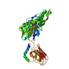 5obuSC 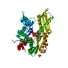 5obwC  5obxC 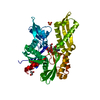 5obyC S: Starting model for refinement C: citing same article ( |
|---|---|
| Similar structure data |


-Links
 PDBj
PDBj
- Assembly
| Deposited unit | 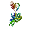
| ||||||||
|---|---|---|---|---|---|---|---|---|---|
| 1 |
| ||||||||
| Unit cell |
|
-Components
| #1: Protein | Mass: 57819.926 Da / Num. of mol.: 1 / Mutation: Polypeptide lacks the last 58 residues. Source method: isolated from a genetically manipulated source Details: Contains a C-terminal hexa-histidine tag. Source: (gene. exp.) Mycoplasma genitalium (strain ATCC 33530 / G-37 / NCTC 10195) (bacteria)Gene: dnaK, hsp70, MG305 / Plasmid: pET21d / Production host: |
|---|---|
| #2: Chemical | ChemComp-ADP /   Mass: 427.201 Da / Num. of mol.: 1 / Source method: obtained synthetically / Formula: C10H15N5O10P2 / Comment: ADP, energy-carrying molecule*YM Mass: 427.201 Da / Num. of mol.: 1 / Source method: obtained synthetically / Formula: C10H15N5O10P2 / Comment: ADP, energy-carrying molecule*YM |
| #3: Chemical | ChemComp-PO4 /   Mass: 94.971 Da / Num. of mol.: 1 / Source method: obtained synthetically / Formula: PO4 Mass: 94.971 Da / Num. of mol.: 1 / Source method: obtained synthetically / Formula: PO4 |
| #4: Water | ChemComp-HOH /  Mass: 18.015 Da / Num. of mol.: 21 / Source method: isolated from a natural source / Formula: H2O Mass: 18.015 Da / Num. of mol.: 21 / Source method: isolated from a natural source / Formula: H2O |
-Experimental details
-Experiment
| Experiment | Method: X-RAY DIFFRACTION / Number of used crystals: 1 |
|---|
- Sample preparation
| Crystal | Density Matthews: 2.52 Å3/Da / Density % sol: 51.14 % |
|---|---|
| Crystal grow | Temperature: 293 K / Method: vapor diffusion, hanging drop / pH: 5.5 / Details: Sodium citrate, PEG MME 5000 and butanol. |
-Data collection
| Diffraction | Mean temperature: 100 K |
|---|---|
| Diffraction source | Source: SYNCHROTRON / Site: ESRF  / Beamline: ID23-1 / Wavelength: 0.9763 Å / Beamline: ID23-1 / Wavelength: 0.9763 Å |
| Detector | Type: DECTRIS PILATUS 6M / Detector: PIXEL / Date: Sep 26, 2013 |
| Radiation | Monochromator: Silicon (111) channel-cut / Protocol: SINGLE WAVELENGTH / Monochromatic (M) / Laue (L): M / Scattering type: x-ray |
| Radiation wavelength | Wavelength: 0.9763 Å / Relative weight: 1 |
| Reflection | Resolution: 2.49→94.43 Å / Num. obs: 20864 / % possible obs: 99.7 % / Redundancy: 7 % / Rmerge(I) obs: 0.05 / Rpim(I) all: 0.016 / Net I/σ(I): 17.2 |
| Reflection shell | Resolution: 2.49→2.63 Å / Redundancy: 6.5 % / Rmerge(I) obs: 0.36 / Mean I/σ(I) obs: 1.2 / Num. unique obs: 2917 / Rpim(I) all: 0.12 / % possible all: 98 |
- Processing
| Software |
| ||||||||||||||||||||||||||||||||||||||||||||||||||||||||||||
|---|---|---|---|---|---|---|---|---|---|---|---|---|---|---|---|---|---|---|---|---|---|---|---|---|---|---|---|---|---|---|---|---|---|---|---|---|---|---|---|---|---|---|---|---|---|---|---|---|---|---|---|---|---|---|---|---|---|---|---|---|---|
| Refinement | Method to determine structure: MOLECULAR REPLACEMENT Starting model: 5OBU Resolution: 2.49→94.43 Å / Cor.coef. Fo:Fc: 0.952 / Cor.coef. Fo:Fc free: 0.925 / SU B: 30.214 / SU ML: 0.297 / Cross valid method: THROUGHOUT / σ(F): 0 / ESU R: 0.575 / ESU R Free: 0.304 Details: U VALUES : WITH TLS ADDED HYDROGENS HAVE BEEN ADDED IN THE RIDING POSITIONS
| ||||||||||||||||||||||||||||||||||||||||||||||||||||||||||||
| Solvent computation | Ion probe radii: 1 Å / Shrinkage radii: 1 Å / VDW probe radii: 1.2 Å | ||||||||||||||||||||||||||||||||||||||||||||||||||||||||||||
| Displacement parameters | Biso max: 113.22 Å2 / Biso mean: 54.721 Å2 / Biso min: 29.2 Å2
| ||||||||||||||||||||||||||||||||||||||||||||||||||||||||||||
| Refinement step | Cycle: final / Resolution: 2.49→94.43 Å
| ||||||||||||||||||||||||||||||||||||||||||||||||||||||||||||
| Refine LS restraints |
| ||||||||||||||||||||||||||||||||||||||||||||||||||||||||||||
| LS refinement shell | Resolution: 2.494→2.559 Å / Rfactor Rfree error: 0 / Total num. of bins used: 20
| ||||||||||||||||||||||||||||||||||||||||||||||||||||||||||||
| Refinement TLS params. | Method: refined / Origin x: 37.673 Å / Origin y: 120.0828 Å / Origin z: 68.9535 Å
|
