[English] 日本語
Yorodumi
- PDB-3ssb: Structure of Insect Metalloproteinase Inhibitor in Complex with T... -

+
Open data


ID or keywords:

Loading...

-
Basic information

Entry
Database: PDB / ID: 3ssb
TitleStructure of Insect Metalloproteinase Inhibitor in Complex with Thermolysin
Components
  • (Inducible metalloproteinase inhibitor ...) x 2
  • Thermolysin
KeywordsHYDROLASE/HYDROLASE INHIBITOR / Thermolysin fold - Family I8 fold / Metalloprotease Thermolysin inhibitor / Zn Binding / Secreted / HYDROLASE-HYDROLASE INHIBITOR complex
Function / homology
Function and homology information


metalloendopeptidase inhibitor activity / thermolysin / anatomical structure morphogenesis / extracellular matrix organization / wound healing / metalloendopeptidase activity / proteolysis / extracellular space / extracellular region / metal ion binding
Similarity search - Function
Trypsin Inhibitor-like, cysteine rich domain / Serine protease inhibitor-like superfamily / Trypsin Inhibitor like cysteine rich domain / Elastase; domain 1 - #10 / Elastase; domain 1 / PepSY domain / Peptidase propeptide and YPEB domain / Peptidase M4, C-terminal / FTP domain / Peptidase M4 domain ...Trypsin Inhibitor-like, cysteine rich domain / Serine protease inhibitor-like superfamily / Trypsin Inhibitor like cysteine rich domain / Elastase; domain 1 - #10 / Elastase; domain 1 / PepSY domain / Peptidase propeptide and YPEB domain / Peptidase M4, C-terminal / FTP domain / Peptidase M4 domain / Peptidase M4 / Thermolysin metallopeptidase, catalytic domain / Thermolysin metallopeptidase, alpha-helical domain / Fungalysin/Thermolysin Propeptide Motif / Neutral Protease Domain 2 / Neutral Protease; domain 2 / Peptidase M4/M1, CTD superfamily / Neutral zinc metallopeptidases, zinc-binding region signature. / Roll / Orthogonal Bundle / Mainly Alpha / Alpha Beta
Similarity search - Domain/homology
Thermolysin / Inducible metalloproteinase inhibitor protein
Similarity search - Component
Biological speciesGalleria mellonella (greater wax moth)
Bacillus thermoproteolyticus (bacteria)
MethodX-RAY DIFFRACTION / SYNCHROTRON / MOLECULAR REPLACEMENT / Resolution: 1.8 Å
AuthorsArolas, J.L. / Botelho, T.O. / Vilcinskas, A. / Gomis-Ruth, F.X.
CitationJournal: Angew.Chem.Int.Ed.Engl. / Year: 2011
Title: Structural evidence for standard-mechanism inhibition in metallopeptidases from a complex poised to resynthesize a Peptide bond.
Authors: Arolas, J.L. / Botelho, T.O. / Vilcinskas, A. / Gomis-Ruth, F.X.
History
DepositionJul 8, 2011Deposition site: RCSB / Processing site: RCSB
Revision 1.0Sep 7, 2011Provider: repository / Type: Initial release
Revision 1.1Nov 2, 2011Group: Database references
Revision 1.2Nov 8, 2017Group: Refinement description / Category: software / Item: _software.name
Revision 1.3Sep 13, 2023Group: Data collection / Database references ...Data collection / Database references / Derived calculations / Refinement description
Category: chem_comp_atom / chem_comp_bond ...chem_comp_atom / chem_comp_bond / database_2 / pdbx_initial_refinement_model / pdbx_struct_conn_angle / struct_conn / struct_ref_seq_dif / struct_site
Item: _database_2.pdbx_DOI / _database_2.pdbx_database_accession ..._database_2.pdbx_DOI / _database_2.pdbx_database_accession / _pdbx_struct_conn_angle.ptnr1_auth_asym_id / _pdbx_struct_conn_angle.ptnr1_auth_comp_id / _pdbx_struct_conn_angle.ptnr1_auth_seq_id / _pdbx_struct_conn_angle.ptnr1_label_asym_id / _pdbx_struct_conn_angle.ptnr1_label_atom_id / _pdbx_struct_conn_angle.ptnr1_label_comp_id / _pdbx_struct_conn_angle.ptnr1_label_seq_id / _pdbx_struct_conn_angle.ptnr2_auth_asym_id / _pdbx_struct_conn_angle.ptnr2_auth_comp_id / _pdbx_struct_conn_angle.ptnr2_auth_seq_id / _pdbx_struct_conn_angle.ptnr2_label_asym_id / _pdbx_struct_conn_angle.ptnr2_label_atom_id / _pdbx_struct_conn_angle.ptnr2_label_comp_id / _pdbx_struct_conn_angle.ptnr3_auth_asym_id / _pdbx_struct_conn_angle.ptnr3_auth_comp_id / _pdbx_struct_conn_angle.ptnr3_auth_seq_id / _pdbx_struct_conn_angle.ptnr3_label_asym_id / _pdbx_struct_conn_angle.ptnr3_label_atom_id / _pdbx_struct_conn_angle.ptnr3_label_comp_id / _pdbx_struct_conn_angle.ptnr3_label_seq_id / _pdbx_struct_conn_angle.value / _struct_conn.pdbx_dist_value / _struct_conn.ptnr1_auth_asym_id / _struct_conn.ptnr1_auth_comp_id / _struct_conn.ptnr1_auth_seq_id / _struct_conn.ptnr1_label_asym_id / _struct_conn.ptnr1_label_atom_id / _struct_conn.ptnr1_label_comp_id / _struct_conn.ptnr1_label_seq_id / _struct_conn.ptnr2_auth_asym_id / _struct_conn.ptnr2_auth_comp_id / _struct_conn.ptnr2_auth_seq_id / _struct_conn.ptnr2_label_asym_id / _struct_conn.ptnr2_label_atom_id / _struct_conn.ptnr2_label_comp_id / _struct_conn.ptnr2_label_seq_id / _struct_ref_seq_dif.details / _struct_site.pdbx_auth_asym_id / _struct_site.pdbx_auth_comp_id / _struct_site.pdbx_auth_seq_id

-
Structure visualization

Structure viewerMolecule:
MolmilJmol/JSmol

Downloads & links

-
Assembly

Deposited unit
A: Thermolysin
C: Inducible metalloproteinase inhibitor protein
I: Inducible metalloproteinase inhibitor protein
B: Thermolysin
D: Inducible metalloproteinase inhibitor protein
J: Inducible metalloproteinase inhibitor protein
hetero molecules


Theoretical massNumber of molelcules
Total (without water)85,35719
Polymers84,6636
Non-polymers69413
Water10,989610
1
A: Thermolysin
C: Inducible metalloproteinase inhibitor protein
I: Inducible metalloproteinase inhibitor protein
hetero molecules


Theoretical massNumber of molelcules
Total (without water)42,72410
Polymers42,3323
Non-polymers3937
Water543
TypeNameSymmetry operationNumber
identity operation1_555x,y,z1
Buried area3440 Å2
ΔGint-21 kcal/mol
Surface area15540 Å2
MethodPISA
2
B: Thermolysin
D: Inducible metalloproteinase inhibitor protein
J: Inducible metalloproteinase inhibitor protein
hetero molecules


Theoretical massNumber of molelcules
Total (without water)42,6329
Polymers42,3323
Non-polymers3016
Water543
TypeNameSymmetry operationNumber
identity operation1_555x,y,z1
Buried area3370 Å2
ΔGint-18 kcal/mol
Surface area15480 Å2
MethodPISA
Unit cell
Length a, b, c (Å)45.640, 78.370, 92.290
Angle α, β, γ (deg.)90.00, 90.08, 90.00
Int Tables number4
Space group name H-MP1211
Details
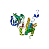
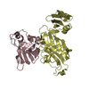
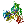
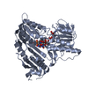
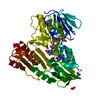
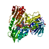
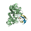
Author states that inhibitor IMPI alpha maintains its 3D structure (represented by chains C,I and D,J) after being cleaved between residues 56-57. The biological assemnbly is a dimeric complex between a proteinase moiety (chains A or B) and an inhibitor moiety (chains C,I or D,J) indicated as trimeric in remark 350.

-
Components

-
Protein , 1 types, 2 molecules AB

#1: Protein Thermolysin / / Thermostable neutral proteinase


Mass: 34360.336 Da / Num. of mol.: 2 / Source method: isolated from a natural source / Source: (natural) Bacillus thermoproteolyticus (bacteria) / References: UniProt: P00800, thermolysin

-
Inducible metalloproteinase inhibitor ... , 2 types, 4 molecules CDIJ

#2: Protein/peptide Inducible metalloproteinase inhibitor protein / IMPI alpha


Mass: 4286.869 Da / Num. of mol.: 2 / Fragment: UNP residues 19-56
Source method: isolated from a genetically manipulated source
Source: (gene. exp.) Galleria mellonella (greater wax moth) / Gene: IMPI / Plasmid: pET-32a / Production host: Escherichia coli (E. coli) / Strain (production host): BL21(DE3) Origami2 / References: UniProt: P82176
#3: Protein/peptide Inducible metalloproteinase inhibitor protein / IMPI alpha


Mass: 3684.299 Da / Num. of mol.: 2 / Fragment: UNP residues 57-88
Source method: isolated from a genetically manipulated source
Source: (gene. exp.) Galleria mellonella (greater wax moth) / Gene: IMPI / Plasmid: pET-32a / Production host: Escherichia coli (E. coli) / Strain (production host): BL21(DE3) Origami2 / References: UniProt: P82176

-
Non-polymers , 5 types, 623 molecules

#4: Chemical ChemComp-GOL / GLYCEROL / GLYCERIN / PROPANE-1,2,3-TRIOL / Glycerol


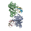
Mass: 92.094 Da / Num. of mol.: 3 / Source method: obtained synthetically / Formula: C3H8O3
#5: Chemical ChemComp-ZN / ZINC ION


Mass: 65.409 Da / Num. of mol.: 2 / Source method: obtained synthetically / Formula: Zn
#6: Chemical
ChemComp-CA / CALCIUM ION


Mass: 40.078 Da / Num. of mol.: 6 / Source method: obtained synthetically / Formula: Ca
#7: Chemical ChemComp-NA / SODIUM ION


Mass: 22.990 Da / Num. of mol.: 2 / Source method: obtained synthetically / Formula: Na
#8: Water ChemComp-HOH / water / Water


Mass: 18.015 Da / Num. of mol.: 610 / Source method: isolated from a natural source / Formula: H2O

-
Experimental details

-
Experiment

ExperimentMethod: X-RAY DIFFRACTION / Number of used crystals: 1

-
Sample preparation

CrystalDensity Matthews: 1.95 Å3/Da / Density % sol: 36.91 %
Crystal growTemperature: 293 K / Method: vapor diffusion, sitting drop / pH: 8.5
Details: 20% w/v PEG 4000, 0.2 M sodium acetate, 0.1 M Tris-HCl, pH 8.5, VAPOR DIFFUSION, SITTING DROP, temperature 293K

-
Data collection

DiffractionMean temperature: 100 K
Diffraction sourceSource: SYNCHROTRON / Site: ESRF / Beamline: ID23-1 / Wavelength: 0.9763 Å
DetectorType: ADSC QUANTUM 315r / Detector: CCD / Date: Nov 25, 2010
RadiationMonochromator: Si(111) / Protocol: SINGLE WAVELENGTH / Monochromatic (M) / Laue (L): M / Scattering type: x-ray
Radiation wavelengthWavelength: 0.9763 Å / Relative weight: 1
ReflectionResolution: 1.8→50 Å / Num. all: 59746 / Num. obs: 59746 / % possible obs: 99.5 % / Observed criterion σ(F): 0 / Observed criterion σ(I): 0 / Rmerge(I) obs: 0.079
Reflection shellResolution: 1.8→1.9 Å / Rmerge(I) obs: 0.079 / Mean I/σ(I) obs: 20.3 / Num. unique all: 59746 / % possible all: 99.5

-
Processing

Software
NameVersionClassification
ProDCdata collection
PHASERphasing
REFMAC5.5.0102refinement
XDSdata reduction
SCALAdata scaling
RefinementMethod to determine structure: MOLECULAR REPLACEMENT
Starting model: PDB ENTRY 2TMN
Resolution: 1.8→50 Å / Cor.coef. Fo:Fc: 0.967 / Cor.coef. Fo:Fc free: 0.956 / SU B: 6.423 / SU ML: 0.089 / Cross valid method: THROUGHOUT / σ(F): 0 / ESU R Free: 0.116 / Stereochemistry target values: MAXIMUM LIKELIHOOD / Details: HYDROGENS HAVE BEEN ADDED IN THE RIDING POSITIONS
RfactorNum. reflection% reflectionSelection details
Rfree0.1931 793 1.3 %RANDOM
Rwork0.15863 ---
all0.15911 59746 --
obs0.15911 58951 99.51 %-
Solvent computationIon probe radii: 0.8 Å / Shrinkage radii: 0.8 Å / VDW probe radii: 1.4 Å / Solvent model: BABINET MODEL WITH MASK
Displacement parametersBiso mean: 28.338 Å2
Baniso -1Baniso -2Baniso -3
1-2.23 Å20 Å20.23 Å2
2---1.06 Å20 Å2
3----1.17 Å2
Refinement stepCycle: LAST / Resolution: 1.8→50 Å
ProteinNucleic acidLigandSolventTotal
Num. atoms5842 0 28 610 6480
Refine LS restraints
Refine-IDTypeDev idealDev ideal targetNumber
X-RAY DIFFRACTIONr_bond_refined_d0.0130.0216031
X-RAY DIFFRACTIONr_bond_other_d0.0010.023860
X-RAY DIFFRACTIONr_angle_refined_deg1.3141.9318167
X-RAY DIFFRACTIONr_angle_other_deg0.90839388
X-RAY DIFFRACTIONr_dihedral_angle_1_deg6.0365752
X-RAY DIFFRACTIONr_dihedral_angle_2_deg34.04924.698298
X-RAY DIFFRACTIONr_dihedral_angle_3_deg12.52715888
X-RAY DIFFRACTIONr_dihedral_angle_4_deg17.6261524
X-RAY DIFFRACTIONr_chiral_restr0.0820.2866
X-RAY DIFFRACTIONr_gen_planes_refined0.0050.026910
X-RAY DIFFRACTIONr_gen_planes_other0.0010.021256
X-RAY DIFFRACTIONr_mcbond_it0.6391.53752
X-RAY DIFFRACTIONr_mcbond_other0.1831.51576
X-RAY DIFFRACTIONr_mcangle_it1.06525979
X-RAY DIFFRACTIONr_scbond_it1.76132279
X-RAY DIFFRACTIONr_scangle_it2.7254.52188
LS refinement shellResolution: 1.803→1.85 Å / Total num. of bins used: 20
RfactorNum. reflection% reflection
Rfree0.284 49 -
Rwork0.207 4150 -
obs--94.81 %
Refinement TLS params.

Method: refined / Refine-ID: X-RAY DIFFRACTION

IDL112)L122)L132)L222)L232)L332)S11 (Å °)S12 (Å °)S13 (Å °)S21 (Å °)S22 (Å °)S23 (Å °)S31 (Å °)S32 (Å °)S33 (Å °)T112)T122)T132)T222)T232)T332)Origin x (Å)Origin y (Å)Origin z (Å)
11.01070.27850.43910.8266-0.32141.4555-0.05160.0185-0.0426-0.0045-0.019-0.1282-0.036-0.03190.07050.0361-0.02040.01980.074-0.02110.147522.20221.50538.712
20.94290.0458-0.71590.28950.28341.67840.0125-0.05620.02940.0419-0.03440.09120.0520.01840.02190.1401-0.0586-0.01070.15240.02460.14581.87717.88684.736
32.71912.04470.83912.8371.44741.10420.17150.185-0.0442-1.145-0.23470.8176-0.0911-0.10430.06310.16530.0618-0.08460.1916-0.00880.22676.060.32933.915
42.71983.413-1.88116.7045-2.20851.66640.1606-0.03910.0984-1.3812-0.2265-0.63670.027-0.01490.06590.31280.02630.10690.21490.01060.182816.67339.58980.018
50.20260.095-0.16820.1736-0.02070.650.0014-0.0144-0.02280.0255-0.014-0.01820.0082-0.01930.01250.1991-0.0116-0.01450.23150.00670.283113.1919.09156.367
Refinement TLS group
IDRefine-IDRefine TLS-IDAuth asym-IDAuth seq-ID
1X-RAY DIFFRACTION1A1 - 316
2X-RAY DIFFRACTION1A999
3X-RAY DIFFRACTION1A998
4X-RAY DIFFRACTION1A997
5X-RAY DIFFRACTION1A996
6X-RAY DIFFRACTION1A995
7X-RAY DIFFRACTION2B1 - 316
8X-RAY DIFFRACTION2B999
9X-RAY DIFFRACTION2B998
10X-RAY DIFFRACTION2B997
11X-RAY DIFFRACTION2B996
12X-RAY DIFFRACTION2B995
13X-RAY DIFFRACTION3I57 - 86
14X-RAY DIFFRACTION3C22 - 56
15X-RAY DIFFRACTION4J57 - 85
16X-RAY DIFFRACTION4D23 - 56
17X-RAY DIFFRACTION5C595 - 1112
18X-RAY DIFFRACTION5D762 - 1101
19X-RAY DIFFRACTION5I501
20X-RAY DIFFRACTION5J853 - 1108

+
About Yorodumi

-
News

-
Feb 9, 2022. New format data for meta-information of EMDB entries

New format data for meta-information of EMDB entries

  • Version 3 of the EMDB header file is now the official format.
  • The previous official version 1.9 will be removed from the archive.

Related info.:EMDB header

External links:wwPDB to switch to version 3 of the EMDB data model

-
Aug 12, 2020. Covid-19 info

Covid-19 info

URL: https://pdbj.org/emnavi/covid19.php

New page: Covid-19 featured information page in EM Navigator.

Related info.:Covid-19 info / Mar 5, 2020. Novel coronavirus structure data

+
Mar 5, 2020. Novel coronavirus structure data

Novel coronavirus structure data

Related info.:Yorodumi Speices / Aug 12, 2020. Covid-19 info

External links:COVID-19 featured content - PDBj / Molecule of the Month (242):Coronavirus Proteases

+
Jan 31, 2019. EMDB accession codes are about to change! (news from PDBe EMDB page)

EMDB accession codes are about to change! (news from PDBe EMDB page)

  • The allocation of 4 digits for EMDB accession codes will soon come to an end. Whilst these codes will remain in use, new EMDB accession codes will include an additional digit and will expand incrementally as the available range of codes is exhausted. The current 4-digit format prefixed with “EMD-” (i.e. EMD-XXXX) will advance to a 5-digit format (i.e. EMD-XXXXX), and so on. It is currently estimated that the 4-digit codes will be depleted around Spring 2019, at which point the 5-digit format will come into force.
  • The EM Navigator/Yorodumi systems omit the EMD- prefix.

Related info.:Q: What is EMD? / ID/Accession-code notation in Yorodumi/EM Navigator

External links:EMDB Accession Codes are Changing Soon! / Contact to PDBj

+
Jul 12, 2017. Major update of PDB

Major update of PDB

  • wwPDB released updated PDB data conforming to the new PDBx/mmCIF dictionary.
  • This is a major update changing the version number from 4 to 5, and with Remediation, in which all the entries are updated.
  • In this update, many items about electron microscopy experimental information are reorganized (e.g. em_software).
  • Now, EM Navigator and Yorodumi are based on the updated data.

External links:wwPDB Remediation / Enriched Model Files Conforming to OneDep Data Standards Now Available in the PDB FTP Archive

-
Yorodumi

Thousand views of thousand structures

  • Yorodumi is a browser for structure data from EMDB, PDB, SASBDB, etc.
  • This page is also the successor to EM Navigator detail page, and also detail information page/front-end page for Omokage search.
  • The word "yorodu" (or yorozu) is an old Japanese word meaning "ten thousand". "mi" (miru) is to see.

Related info.:EMDB / PDB / SASBDB / Comparison of 3 databanks / Yorodumi Search / Aug 31, 2016. New EM Navigator & Yorodumi / Yorodumi Papers / Jmol/JSmol / Function and homology information / Changes in new EM Navigator and Yorodumi

Read more