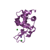
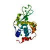
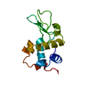
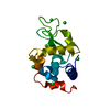
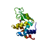
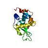
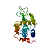
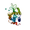
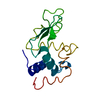
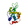
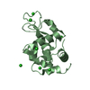
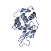
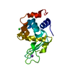
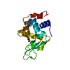
ジャーナル: Acta Crystallogr D Struct Biol / 年: 2017 タイトル: Protein structure determination by electron diffraction using a single three-dimensional nanocrystal. 著者: M T B Clabbers / E van Genderen / W Wan / E L Wiegers / T Gruene / J P Abrahams / 要旨: Three-dimensional nanometre-sized crystals of macromolecules currently resist structure elucidation by single-crystal X-ray crystallography. Here, a single nanocrystal with a diffracting volume of ...Three-dimensional nanometre-sized crystals of macromolecules currently resist structure elucidation by single-crystal X-ray crystallography. Here, a single nanocrystal with a diffracting volume of only 0.14 µm, i.e. no more than 6 × 10 unit cells, provided sufficient information to determine the structure of a rare dimeric polymorph of hen egg-white lysozyme by electron crystallography. This is at least an order of magnitude smaller than was previously possible. The molecular-replacement solution, based on a monomeric polyalanine model, provided sufficient phasing power to show side-chain density, and automated model building was used to reconstruct the side chains. Diffraction data were acquired using the rotation method with parallel beam diffraction on a Titan Krios transmission electron microscope equipped with a novel in-house-designed 1024 × 1024 pixel Timepix hybrid pixel detector for low-dose diffraction data collection. Favourable detector characteristics include the ability to accurately discriminate single high-energy electrons from X-rays and count them, fast readout to finely sample reciprocal space and a high dynamic range. This work, together with other recent milestones, suggests that electron crystallography can provide an attractive alternative in determining biological structures.
詳細: Parameters where extreme values were added are not applicable to our experiment and can not be filled out because there is no experimental data, these include Rsym, overall phase error, ...詳細: Parameters where extreme values were added are not applicable to our experiment and can not be filled out because there is no experimental data, these include Rsym, overall phase error, overall phase residual and phase error rejection フーリエ空間範囲: 61.24 % / 再高解像度: 2.11 Å / 測定した強度の数: 41191 / 構造因子数: 8503 / 位相誤差: 179.9 ° / 位相残差: 179.9 ° / 位相誤差の除外基準: 9999 / Rmerge: 39.8 / Rsym: 100
-
解析
ソフトウェア
名称: REFMAC / バージョン: 5.8.0158 / 分類: 精密化
EM 3D crystal entity
∠α: 90 ° / ∠β: 90 ° / ∠γ: 90 ° / A: 104.56 Å / B: 68.05 Å / C: 32.05 Å / 空間群名: P21212 / 空間群番号: 18
CTF補正
タイプ: NONE
3次元再構成
解像度の算出法: DIFFRACTION PATTERN/LAYERLINES / 対称性のタイプ: 3D CRYSTAL
解像度: 2.11→57.03 Å / Cor.coef. Fo:Fc: 0.905 / SU B: 10.574 / SU ML: 0.263 / 交差検証法: NONE / ESU R: 1.306 詳細: We present Rcomplete instead of Rfree, calculating Rcomplete takes into account all reflections which is preferred when considering data sets with less than about 10,000 unique reflections ...詳細: We present Rcomplete instead of Rfree, calculating Rcomplete takes into account all reflections which is preferred when considering data sets with less than about 10,000 unique reflections [Brunger (1997) Methods in Enzymology 277:366-396, Luebben & Gruene (2015) PNAS 112:8999-9003]. To calculate Rcomplete a 0.2% test set size was chosen i.e. 500 test sets were created. First all non-measured observations were removed from the reflection file, and the 500 test sets were assigned at random. The model was then refined independently each time omitting one of the different test sets, thus in total 500 iterations. Each time refinement was carried out until practically reaching convergence. Finally, the value for Rcomplete was calculated from the excluded data i.e. calculated from all reflections. Since all structure factors are used in turn this leads to a more robust calculation than Rfree.
 ムービー
ムービー コントローラー


 データを開く
 基本情報
基本情報 要素
要素 キーワード
キーワード 機能・相同性情報
 分子置換 / クライオ電子顕微鏡法 / 解像度: 2.11 Å
  データ登録者
 引用
引用


 構造の表示
構造の表示 ダウンロードとリンク
 その他のダウンロード


 PDBj
PDBj


 集合体
集合体
 試料調製
試料調製
 解析
解析