 Movie
Movie Controller

[English] 日本語
 Yorodumi
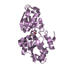
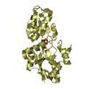
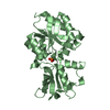
- PDB-5o2j: Pseudomonas stutzeri PtxB in complex with phosphite to 1.52 A res... -
+ Open data

- Basic information
| Entry | Database: PDB / ID: 5o2j | ||||||||||||||||||
|---|---|---|---|---|---|---|---|---|---|---|---|---|---|---|---|---|---|---|---|
| Title | Pseudomonas stutzeri PtxB in complex with phosphite to 1.52 A resolution | ||||||||||||||||||
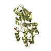
 Components Components | Probable phosphite transport system-binding protein PtxB | ||||||||||||||||||
 Keywords Keywords | TRANSPORT PROTEIN / phosphite / ABC transporters / marine bacteria / PBP / SBP | ||||||||||||||||||
| Function / homology |  Function and homology information Function and homology informationATP-binding cassette (ABC) transporter complex / transmembrane transport Similarity search - Function | ||||||||||||||||||
| Biological species |  Pseudomonas stutzeri (bacteria) Pseudomonas stutzeri (bacteria) | ||||||||||||||||||
| Method |  X-RAY DIFFRACTION / SYNCHROTRON / MOLECULAR REPLACEMENT / Resolution: 1.52 Å X-RAY DIFFRACTION / SYNCHROTRON / MOLECULAR REPLACEMENT / Resolution: 1.52 Å | ||||||||||||||||||
 Authors Authors | Bisson, C. / Hitchcock, A. | ||||||||||||||||||
| Funding support |  United Kingdom, 5items United Kingdom, 5items
| ||||||||||||||||||
 Citation Citation | Journal: Nat Commun / Year: 2017 Title: The molecular basis of phosphite and hypophosphite recognition by ABC-transporters. Authors: Bisson, C. / Adams, N.B.P. / Stevenson, B. / Brindley, A.A. / Polyviou, D. / Bibby, T.S. / Baker, P.J. / Hunter, C.N. / Hitchcock, A. | ||||||||||||||||||
| History |
|
- Structure visualization
| Structure viewer | Molecule: MolmilJmol/JSmol |
|---|
- Downloads & links
-Download
| PDBx/mmCIF format | 5o2j.cif.gz | 68.4 KB | Display | PDBx/mmCIF format |
|---|---|---|---|---|
| PDB format | pdb5o2j.ent.gz | 49.3 KB | Display | PDB format |
| PDBx/mmJSON format | 5o2j.json.gz | Tree view | PDBx/mmJSON format | |
| Others |  Other downloads Other downloads |
-Validation report
| Arichive directory | https://data.pdbj.org/pub/pdb/validation_reports/o2/5o2jftp://data.pdbj.org/pub/pdb/validation_reports/o2/5o2j | HTTPS FTP |
|---|
-Related structure data
| Related structure data |  5jvbC 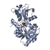 5lq1C 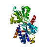 5lq5C 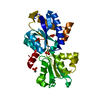 5lq8C 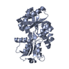 5lv1C  5me4C  5o2kC  5o37C C: citing same article ( |
|---|---|
| Similar structure data |


-Links
 PDBj
PDBj- Assembly
| Deposited unit | 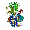
| ||||||||
|---|---|---|---|---|---|---|---|---|---|
| 1 |
| ||||||||
| Unit cell |
|
-Components
| #1: Protein | Mass: 30173.084 Da / Num. of mol.: 1 Source method: isolated from a genetically manipulated source Details: 6x His-tag at C-terminus / Source: (gene. exp.) Pseudomonas stutzeri (bacteria) / Gene: ptxB / Production host: |
|---|---|
| #2: Chemical | ChemComp-2PO / 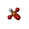  Mass: 79.980 Da / Num. of mol.: 1 / Source method: obtained synthetically / Formula: HO3P Mass: 79.980 Da / Num. of mol.: 1 / Source method: obtained synthetically / Formula: HO3P |
| #3: Chemical | ChemComp-EDO /   Mass: 62.068 Da / Num. of mol.: 1 / Source method: obtained synthetically / Formula: C2H6O2 Mass: 62.068 Da / Num. of mol.: 1 / Source method: obtained synthetically / Formula: C2H6O2 |
| #4: Water | ChemComp-HOH /  Mass: 18.015 Da / Num. of mol.: 193 / Source method: isolated from a natural source / Formula: H2O Mass: 18.015 Da / Num. of mol.: 193 / Source method: isolated from a natural source / Formula: H2O |
-Experimental details
-Experiment
| Experiment | Method: X-RAY DIFFRACTION / Number of used crystals: 1 |
|---|
- Sample preparation
| Crystal | Density Matthews: 2.33 Å3/Da / Density % sol: 47.11 % / Description: rods |
|---|---|
| Crystal grow | Temperature: 290 K / Method: vapor diffusion, sitting drop / pH: 5 / Details: 0.1 M MMT buffer pH 5 25% (w/v) and PEG 1500 |
-Data collection
| Diffraction | Mean temperature: 100 K |
|---|---|
| Diffraction source | Source: SYNCHROTRON / Site: Diamond / Beamline: I04-1 / Wavelength: 0.92819 Å |
| Detector | Type: DECTRIS PILATUS 6M / Detector: PIXEL / Date: Feb 5, 2017 |
| Radiation | Protocol: SINGLE WAVELENGTH / Monochromatic (M) / Laue (L): M / Scattering type: x-ray |
| Radiation wavelength | Wavelength: 0.92819 Å / Relative weight: 1 |
| Reflection | Resolution: 1.52→33.25 Å / Num. obs: 44234 / % possible obs: 100 % / Redundancy: 12.4 % / CC1/2: 0.999 / Rpim(I) all: 0.039 / Net I/σ(I): 11.9 |
| Reflection shell | Resolution: 1.52→1.55 Å / Redundancy: 10.3 % / Mean I/σ(I) obs: 1.2 / Num. unique obs: 2169 / CC1/2: 0.51 / Rpim(I) all: 0.607 / % possible all: 100 |
- Processing
| Software |
| ||||||||||||||||||||||||||||||||||||||||||||||||||||||||||||||||||||||||||||||||||||||||||||||||||||||||||||||||||||||||||||||||||||||||||||||||||||||||||||||||||||||||||||||||||||||
|---|---|---|---|---|---|---|---|---|---|---|---|---|---|---|---|---|---|---|---|---|---|---|---|---|---|---|---|---|---|---|---|---|---|---|---|---|---|---|---|---|---|---|---|---|---|---|---|---|---|---|---|---|---|---|---|---|---|---|---|---|---|---|---|---|---|---|---|---|---|---|---|---|---|---|---|---|---|---|---|---|---|---|---|---|---|---|---|---|---|---|---|---|---|---|---|---|---|---|---|---|---|---|---|---|---|---|---|---|---|---|---|---|---|---|---|---|---|---|---|---|---|---|---|---|---|---|---|---|---|---|---|---|---|---|---|---|---|---|---|---|---|---|---|---|---|---|---|---|---|---|---|---|---|---|---|---|---|---|---|---|---|---|---|---|---|---|---|---|---|---|---|---|---|---|---|---|---|---|---|---|---|---|---|
| Refinement | Method to determine structure: MOLECULAR REPLACEMENT / Resolution: 1.52→33.25 Å / Cor.coef. Fo:Fc: 0.973 / Cor.coef. Fo:Fc free: 0.955 / SU B: 1.252 / SU ML: 0.044 / Cross valid method: THROUGHOUT / ESU R: 0.063 / ESU R Free: 0.067 / Details: HYDROGENS HAVE BEEN ADDED IN THE RIDING POSITIONS
| ||||||||||||||||||||||||||||||||||||||||||||||||||||||||||||||||||||||||||||||||||||||||||||||||||||||||||||||||||||||||||||||||||||||||||||||||||||||||||||||||||||||||||||||||||||||
| Solvent computation | Ion probe radii: 0.8 Å / Shrinkage radii: 0.8 Å / VDW probe radii: 1.2 Å | ||||||||||||||||||||||||||||||||||||||||||||||||||||||||||||||||||||||||||||||||||||||||||||||||||||||||||||||||||||||||||||||||||||||||||||||||||||||||||||||||||||||||||||||||||||||
| Displacement parameters | Biso mean: 22.051 Å2
| ||||||||||||||||||||||||||||||||||||||||||||||||||||||||||||||||||||||||||||||||||||||||||||||||||||||||||||||||||||||||||||||||||||||||||||||||||||||||||||||||||||||||||||||||||||||
| Refinement step | Cycle: 1 / Resolution: 1.52→33.25 Å
| ||||||||||||||||||||||||||||||||||||||||||||||||||||||||||||||||||||||||||||||||||||||||||||||||||||||||||||||||||||||||||||||||||||||||||||||||||||||||||||||||||||||||||||||||||||||
| Refine LS restraints |
|