 Movie
Movie Controller

[English] 日本語
 Yorodumi
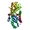
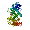
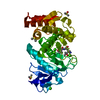
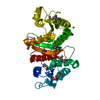
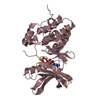
- PDB-5lq5: 1.46 A resolution structure of PhnD1 from Prochlorococcus marinus... -
+ Open data

- Basic information
| Entry | Database: PDB / ID: 5lq5 | |||||||||||||||
|---|---|---|---|---|---|---|---|---|---|---|---|---|---|---|---|---|
| Title | 1.46 A resolution structure of PhnD1 from Prochlorococcus marinus (MIT 9301) in complex with phosphite | |||||||||||||||
 Components Components | Putative phosphonate binding protein for ABC transporter | |||||||||||||||
 Keywords Keywords | TRANSPORT PROTEIN / ABC-transporter / phosphite / Prochlorococcus / periplasmic binding protein | |||||||||||||||
| Function / homology | Putative ABC transporter periplasmic binding protein PhnD-like / Phosphate/phosphite/phosphonate ABC transporter, periplasmic binding protein / ABC transporter, phosphonate, periplasmic substrate-binding protein / ATP-binding cassette (ABC) transporter complex / transmembrane transport / periplasmic space / PHOSPHITE ION / Probable ABC transporter phosphite binding protein PhnD1 Function and homology information Function and homology information | |||||||||||||||
| Biological species |  Prochlorococcus marinus str. MIT 9301 (bacteria) Prochlorococcus marinus str. MIT 9301 (bacteria) | |||||||||||||||
| Method |  X-RAY DIFFRACTION / SYNCHROTRON / MOLECULAR REPLACEMENT / Resolution: 1.46 Å X-RAY DIFFRACTION / SYNCHROTRON / MOLECULAR REPLACEMENT / Resolution: 1.46 Å | |||||||||||||||
 Authors Authors | Bisson, C. / Adams, N.B.P. / Polyviou, D. / Bibby, T.S. / Hunter, C.N. / Hitchcock, A. | |||||||||||||||
| Funding support |  United Kingdom, 4items United Kingdom, 4items
| |||||||||||||||
 Citation Citation | Journal: Nat Commun / Year: 2017 Title: The molecular basis of phosphite and hypophosphite recognition by ABC-transporters. Authors: Bisson, C. / Adams, N.B.P. / Stevenson, B. / Brindley, A.A. / Polyviou, D. / Bibby, T.S. / Baker, P.J. / Hunter, C.N. / Hitchcock, A. | |||||||||||||||
| History |
|
- Structure visualization
| Structure viewer | Molecule: MolmilJmol/JSmol |
|---|
- Downloads & links
-Download
| PDBx/mmCIF format | 5lq5.cif.gz | 130.2 KB | Display | PDBx/mmCIF format |
|---|---|---|---|---|
| PDB format | pdb5lq5.ent.gz | 101.9 KB | Display | PDB format |
| PDBx/mmJSON format | 5lq5.json.gz | Tree view | PDBx/mmJSON format | |
| Others |  Other downloads Other downloads |
-Validation report
| Arichive directory | https://data.pdbj.org/pub/pdb/validation_reports/lq/5lq5ftp://data.pdbj.org/pub/pdb/validation_reports/lq/5lq5 | HTTPS FTP |
|---|
-Related structure data
| Related structure data |  5jvbSC  5lq1C  5lq8C  5lv1C  5me4C  5o2jC  5o2kC  5o37C C: citing same article ( S: Starting model for refinement |
|---|---|
| Similar structure data |


-Links
 PDBj
PDBj- Assembly
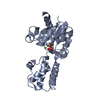
| Deposited unit | 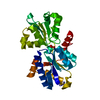
| ||||||||
|---|---|---|---|---|---|---|---|---|---|
| 1 |
| ||||||||
| Unit cell |
|
-Components
| #1: Protein | Mass: 32329.049 Da / Num. of mol.: 1 Source method: isolated from a genetically manipulated source Details: The construct has been truncated by 24 residues on the N-terminus to remove a signal peptide and has a C-terminal 6XHis tag. Source: (gene. exp.) Prochlorococcus marinus str. MIT 9301 (bacteria)Gene: phnD, P9301_07261 / Production host: |
|---|---|
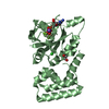
| #2: Chemical | ChemComp-PO3 / 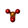  Mass: 78.972 Da / Num. of mol.: 1 / Source method: obtained synthetically / Formula: PO3 Mass: 78.972 Da / Num. of mol.: 1 / Source method: obtained synthetically / Formula: PO3 |
| #3: Water | ChemComp-HOH /  Mass: 18.015 Da / Num. of mol.: 129 / Source method: isolated from a natural source / Formula: H2O Mass: 18.015 Da / Num. of mol.: 129 / Source method: isolated from a natural source / Formula: H2O |
-Experimental details
-Experiment
| Experiment | Method: X-RAY DIFFRACTION / Number of used crystals: 1 |
|---|
- Sample preparation
| Crystal | Density Matthews: 2.16 Å3/Da / Density % sol: 43.09 % |
|---|---|
| Crystal grow | Temperature: 290 K / Method: vapor diffusion, sitting drop / pH: 8 Details: 0.1 M SPG (succinic acid, sodium dihydrogen phosphate, glycine) buffer pH 8.0 and 25% (w/v) PEG 1500 |
-Data collection
| Diffraction | Mean temperature: 100 K |
|---|---|
| Diffraction source | Source: SYNCHROTRON / Site: Diamond / Beamline: I24 / Wavelength: 0.97951 Å |
| Detector | Type: DECTRIS PILATUS 6M / Detector: PIXEL / Date: Jul 1, 2016 |
| Radiation | Protocol: SINGLE WAVELENGTH / Monochromatic (M) / Laue (L): M / Scattering type: x-ray |
| Radiation wavelength | Wavelength: 0.97951 Å / Relative weight: 1 |
| Reflection | Resolution: 1.46→44.27 Å / Num. obs: 46137 / % possible obs: 97.3 % / Redundancy: 3.5 % / CC1/2: 0.999 / Rmerge(I) obs: 0.021 / Net I/σ(I): 15.8 |
| Reflection shell | Resolution: 1.46→1.49 Å / Redundancy: 2.5 % / Rmerge(I) obs: 0.67 / Mean I/σ(I) obs: 1.1 / CC1/2: 0.569 / % possible all: 78.4 |
- Processing
| Software |
| ||||||||||||||||||||||||||||||||||||||||||||||||||||||||||||||||||||||||||||||||||||||||||||||||||||||||||||||||||||||||||||||||||||||||||||||||||||||||||||||||||||||||||||||||||||||
|---|---|---|---|---|---|---|---|---|---|---|---|---|---|---|---|---|---|---|---|---|---|---|---|---|---|---|---|---|---|---|---|---|---|---|---|---|---|---|---|---|---|---|---|---|---|---|---|---|---|---|---|---|---|---|---|---|---|---|---|---|---|---|---|---|---|---|---|---|---|---|---|---|---|---|---|---|---|---|---|---|---|---|---|---|---|---|---|---|---|---|---|---|---|---|---|---|---|---|---|---|---|---|---|---|---|---|---|---|---|---|---|---|---|---|---|---|---|---|---|---|---|---|---|---|---|---|---|---|---|---|---|---|---|---|---|---|---|---|---|---|---|---|---|---|---|---|---|---|---|---|---|---|---|---|---|---|---|---|---|---|---|---|---|---|---|---|---|---|---|---|---|---|---|---|---|---|---|---|---|---|---|---|---|
| Refinement | Method to determine structure: MOLECULAR REPLACEMENT Starting model: 5JVB Resolution: 1.46→44.27 Å / Cor.coef. Fo:Fc: 0.98 / Cor.coef. Fo:Fc free: 0.974 / SU B: 2.675 / SU ML: 0.044 / Cross valid method: THROUGHOUT / ESU R: 0.07 / ESU R Free: 0.062 / Stereochemistry target values: MAXIMUM LIKELIHOOD / Details: HYDROGENS HAVE BEEN ADDED IN THE RIDING POSITIONS
| ||||||||||||||||||||||||||||||||||||||||||||||||||||||||||||||||||||||||||||||||||||||||||||||||||||||||||||||||||||||||||||||||||||||||||||||||||||||||||||||||||||||||||||||||||||||
| Solvent computation | Ion probe radii: 0.8 Å / Shrinkage radii: 0.8 Å / VDW probe radii: 1.2 Å / Solvent model: MASK | ||||||||||||||||||||||||||||||||||||||||||||||||||||||||||||||||||||||||||||||||||||||||||||||||||||||||||||||||||||||||||||||||||||||||||||||||||||||||||||||||||||||||||||||||||||||
| Displacement parameters | Biso mean: 34.119 Å2
| ||||||||||||||||||||||||||||||||||||||||||||||||||||||||||||||||||||||||||||||||||||||||||||||||||||||||||||||||||||||||||||||||||||||||||||||||||||||||||||||||||||||||||||||||||||||
| Refinement step | Cycle: LAST / Resolution: 1.46→44.27 Å
| ||||||||||||||||||||||||||||||||||||||||||||||||||||||||||||||||||||||||||||||||||||||||||||||||||||||||||||||||||||||||||||||||||||||||||||||||||||||||||||||||||||||||||||||||||||||
| Refine LS restraints |
|