 Movie
Movie Controller


+ Open data

- Basic information
| Entry | Database: PDB / ID: 6h3v | |||||||||
|---|---|---|---|---|---|---|---|---|---|---|
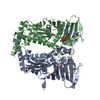
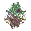
| Title | Bunyamwera Virus Glycoprotein Gc Head Domain | |||||||||
 Components Components | Envelopment polyprotein | |||||||||
 Keywords Keywords | VIRAL PROTEIN / Envelope Glycoprotein | |||||||||
| Function / homology |  Function and homology information Function and homology information: / host cell Golgi membrane / host cell endoplasmic reticulum membrane / fusion of virus membrane with host endosome membrane / symbiont entry into host cell / virion attachment to host cell / virion membrane / membrane Similarity search - Function | |||||||||
| Biological species |  Bunyamwera virus Bunyamwera virus | |||||||||
| Method |  X-RAY DIFFRACTION / SYNCHROTRON / MOLECULAR REPLACEMENT / Resolution: 2.9 Å X-RAY DIFFRACTION / SYNCHROTRON / MOLECULAR REPLACEMENT / Resolution: 2.9 Å | |||||||||
 Authors Authors | Hellert, J. / Aebischer, A. / Wernike, K. / Haouz, A. / Brocchi, E. / Reiche, S. / Guardado-Calvo, P. / Beer, M. / Rey, F.A. | |||||||||
| Funding support | 1items
| |||||||||
 Citation Citation | Journal: Nat Commun / Year: 2019 Title: Orthobunyavirus spike architecture and recognition by neutralizing antibodies. Authors: Hellert, J. / Aebischer, A. / Wernike, K. / Haouz, A. / Brocchi, E. / Reiche, S. / Guardado-Calvo, P. / Beer, M. / Rey, F.A. | |||||||||
| History |
|
- Structure visualization
| Structure viewer | Molecule: MolmilJmol/JSmol |
|---|
- Downloads & links
-Download
| PDBx/mmCIF format | 6h3v.cif.gz | 118.6 KB | Display | PDBx/mmCIF format |
|---|---|---|---|---|
| PDB format | pdb6h3v.ent.gz | 90.8 KB | Display | PDB format |
| PDBx/mmJSON format | 6h3v.json.gz | Tree view | PDBx/mmJSON format | |
| Others |  Other downloads Other downloads |
-Validation report
| Arichive directory | https://data.pdbj.org/pub/pdb/validation_reports/h3/6h3vftp://data.pdbj.org/pub/pdb/validation_reports/h3/6h3v | HTTPS FTP |
|---|
-Related structure data
| Related structure data |  6h3sC 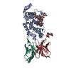 6h3tC  6h3uC 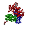 6h3wSC  6h3xC S: Starting model for refinement C: citing same article ( |
|---|---|
| Similar structure data |

-Links
 PDBj
PDBj- Assembly
| Deposited unit | 
| ||||||||
|---|---|---|---|---|---|---|---|---|---|
| 1 | 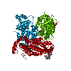
| ||||||||
| Unit cell |
|
-Components
| #1: Protein | Mass: 29526.775 Da / Num. of mol.: 1 / Fragment: Glycoprotein Gc Head Domain Source method: isolated from a genetically manipulated source Source: (gene. exp.) Bunyamwera virus / Gene: GP / Production host:  |
|---|---|
| #2: Polysaccharide | 2-acetamido-2-deoxy-beta-D-glucopyranose-(1-4)-[alpha-L-fucopyranose-(1-6)]2-acetamido-2-deoxy-beta- ...2-acetamido-2-deoxy-beta-D-glucopyranose-(1-4)-[alpha-L-fucopyranose-(1-6)]2-acetamido-2-deoxy-beta-D-glucopyranose Source method: isolated from a genetically manipulated source |
| #3: Chemical | ChemComp-CL /   Mass: 35.453 Da / Num. of mol.: 1 / Source method: obtained synthetically / Formula: Cl Mass: 35.453 Da / Num. of mol.: 1 / Source method: obtained synthetically / Formula: Cl |
| Has protein modification | Y |
-Experimental details
-Experiment
| Experiment | Method: X-RAY DIFFRACTION / Number of used crystals: 1 |
|---|
- Sample preparation
| Crystal | Density Matthews: 3.79 Å3/Da / Density % sol: 67.51 % |
|---|---|
| Crystal grow | Temperature: 291.15 K / Method: vapor diffusion, sitting drop Details: 0.2 uL of 22.2 mg/mL BUNV Gc head domain in 20 mM Tris-Cl pH 8.0, 150 mM NaCl were added to 0.2 uL of reservoir solution containing 0.2 M sodium acetate, 0.1 M Tris-Cl pH 8.5, 30% w/v PEG 4K |
-Data collection
| Diffraction | Mean temperature: 100 K |
|---|---|
| Diffraction source | Source: SYNCHROTRON / Site: ESRF  / Beamline: ID30B / Wavelength: 1.033199 Å / Beamline: ID30B / Wavelength: 1.033199 Å |
| Detector | Type: DECTRIS PILATUS 6M / Detector: PIXEL / Date: Feb 2, 2017 |
| Radiation | Protocol: SINGLE WAVELENGTH / Monochromatic (M) / Laue (L): M / Scattering type: x-ray |
| Radiation wavelength | Wavelength: 1.033199 Å / Relative weight: 1 |
| Reflection | Resolution: 2.9→46.55 Å / Num. obs: 9534 / % possible obs: 99.8 % / Redundancy: 9.6 % / Biso Wilson estimate: 82 Å2 / CC1/2: 1 / Rmerge(I) obs: 0.09 / Rpim(I) all: 0.03 / Rrim(I) all: 0.09 / Net I/σ(I): 16.1 |
| Reflection shell | Resolution: 2.9→3.08 Å / Redundancy: 10 % / Rmerge(I) obs: 1.27 / Mean I/σ(I) obs: 1.9 / Num. unique obs: 1519 / CC1/2: 0.7 / Rpim(I) all: 0.42 / Rrim(I) all: 1.34 / % possible all: 99.8 |
- Processing
| Software |
| ||||||||||||||||||||||||||||||||||||||||||||||||
|---|---|---|---|---|---|---|---|---|---|---|---|---|---|---|---|---|---|---|---|---|---|---|---|---|---|---|---|---|---|---|---|---|---|---|---|---|---|---|---|---|---|---|---|---|---|---|---|---|---|
| Refinement | Method to determine structure: MOLECULAR REPLACEMENT Starting model: 6H3W Resolution: 2.9→46.55 Å / SU ML: 0.39 / Cross valid method: THROUGHOUT / σ(F): 1.35 / Phase error: 23.74
| ||||||||||||||||||||||||||||||||||||||||||||||||
| Solvent computation | Shrinkage radii: 0.9 Å / VDW probe radii: 1.11 Å | ||||||||||||||||||||||||||||||||||||||||||||||||
| Displacement parameters | Biso max: 237.71 Å2 / Biso mean: 103.2644 Å2 / Biso min: 52.84 Å2 | ||||||||||||||||||||||||||||||||||||||||||||||||
| Refinement step | Cycle: final / Resolution: 2.9→46.55 Å
| ||||||||||||||||||||||||||||||||||||||||||||||||
| Refine LS restraints |
| ||||||||||||||||||||||||||||||||||||||||||||||||
| LS refinement shell | Refine-ID: X-RAY DIFFRACTION / Rfactor Rfree error: 0 / Total num. of bins used: 7 / % reflection obs: 100 %
| ||||||||||||||||||||||||||||||||||||||||||||||||
| Refinement TLS params. | Method: refined / Origin x: 62.4934 Å / Origin y: -16.19 Å / Origin z: 19.9856 Å
| ||||||||||||||||||||||||||||||||||||||||||||||||
| Refinement TLS group | Selection details: (chain 'A' and resid 478 through 803) |
