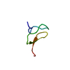
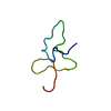
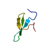
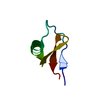
登録情報 データベース : PDB / ID : 5o0uタイトル Crystal structure of tarantula venom peptide Protoxin-II Beta/omega-theraphotoxin-Tp2a キーワード / / / 機能・相同性 / / / / / 生物種 Thrixopelma pruriens (クモ)手法 / / / 解像度 : 0.99 Å データ登録者 Tabor, A. / McCarthy, S. / Reyes, F.E. 資金援助 組織 認可番号 国 Wellcome Trust 109073/Z/15/Z
ジャーナル : J.Am.Chem.Soc. / 年 : 2017タイトル : The Role of Disulfide Bond Replacements in Analogues of the Tarantula Toxin ProTx-II and Their Effects on Inhibition of the Voltage-Gated Sodium Ion Channel Nav1.7.著者 : Wright, Z.V.F. / McCarthy, S. / Dickman, R. / Reyes, F.E. / Sanchez-Martinez, S. / Cryar, A. / Kilford, I. / Hall, A. / Takle, A.K. / Topf, M. / Gonen, T. / Thalassinos, K. / Tabor, A.B. 履歴 登録 2017年5月17日 登録サイト / 処理サイト 改定 1.0 2017年9月13日 Provider / タイプ 改定 1.1 2019年3月27日 Group / Database referencesカテゴリ / citation_author / pdbx_database_procItem _citation.country / _citation.journal_abbrev ... _citation.country / _citation.journal_abbrev / _citation.journal_id_ASTM / _citation.journal_id_CSD / _citation.journal_id_ISSN / _citation.journal_volume / _citation.page_first / _citation.page_last / _citation.pdbx_database_id_DOI / _citation.pdbx_database_id_PubMed / _citation.title / _citation.year 改定 1.2 2024年10月16日 Group / Database references / Structure summaryカテゴリ chem_comp_atom / chem_comp_bond ... chem_comp_atom / chem_comp_bond / database_2 / pdbx_entry_details / pdbx_modification_feature Item / _database_2.pdbx_database_accession
すべて表示 表示を減らす
 ムービー
ムービー コントローラー


 データを開く
 基本情報
基本情報 要素
要素 キーワード
キーワード 機能・相同性情報
 Thrixopelma pruriens (クモ)
 X線回折 /
  データ登録者
 英国, 1件
  引用
引用 構造の表示
構造の表示 ダウンロードとリンク
 その他のダウンロード
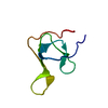


 PDBj
PDBj 集合体
集合体

 分子量: 35.453 Da / 分子数: 2 / 由来タイプ: 合成 / 式: Cl
 分子量: 62.068 Da / 分子数: 2 / 由来タイプ: 合成 / 式: C2H6O2
 分子量: 18.015 Da / 分子数: 54 / 由来タイプ: 天然 / 式: H2O
 試料調製
試料調製 / ビームライン: 5.0.2 / 波長: 1 Å
 解析
解析