 Movie
Movie Controller


+ Open data

- Basic information
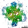
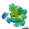
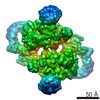
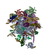
| Entry | Database: PDB / ID: 5o09 | ||||||
|---|---|---|---|---|---|---|---|
| Title | BtubABC mini microtubule | ||||||
 Components Components |
| ||||||
 Keywords Keywords | STRUCTURAL PROTEIN / bacterial cytoskeleton / microtubules | ||||||
| Function / homology |  Function and homology information Function and homology informationmicrotubule-based process / structural constituent of cytoskeleton / microtubule / hydrolase activity / protein serine/threonine kinase activity / GTPase activity / GTP binding Similarity search - Function | ||||||
| Biological species |  Prosthecobacter dejongeii (bacteria)Prosthecobacter vanneervenii (bacteria) Prosthecobacter dejongeii (bacteria)Prosthecobacter vanneervenii (bacteria) | ||||||
| Method | ELECTRON MICROSCOPY / helical reconstruction / cryo EM / Resolution: 3.6 Å | ||||||
 Authors Authors | Deng, X. / Bharat, T.A.M. / Lowe, J. | ||||||
 Citation Citation | Journal: Proc Natl Acad Sci U S A / Year: 2017 Title: Four-stranded mini microtubules formed by BtubAB show dynamic instability. Authors: Xian Deng / Gero Fink / Tanmay A M Bharat / Shaoda He / Danguole Kureisaite-Ciziene / Jan Löwe /  Abstract: Microtubules, the dynamic, yet stiff hollow tubes built from αβ-tubulin protein heterodimers, are thought to be present only in eukaryotic cells. Here, we report a 3.6-Å helical reconstruction ...Microtubules, the dynamic, yet stiff hollow tubes built from αβ-tubulin protein heterodimers, are thought to be present only in eukaryotic cells. Here, we report a 3.6-Å helical reconstruction electron cryomicroscopy structure of four-stranded mini microtubules formed by bacterial tubulin-like BtubAB proteins. Despite their much smaller diameter, mini microtubules share many key structural features with eukaryotic microtubules, such as an M-loop, alternating subunits, and a seam that breaks overall helical symmetry. Using in vitro total internal reflection fluorescence microscopy, we show that bacterial mini microtubules treadmill and display dynamic instability, another hallmark of eukaryotic microtubules. The third protein in the gene cluster, BtubC, previously known as "bacterial kinesin light chain," binds along protofilaments every 8 nm, inhibits BtubAB mini microtubule catastrophe, and increases rescue. Our work reveals that some bacteria contain regulated and dynamic cytomotive microtubule systems that were once thought to be only useful in much larger and sophisticated eukaryotic cells. | ||||||
| History |
|
- Structure visualization
| Movie |
Movie viewer |
|---|---|
| Structure viewer | Molecule: MolmilJmol/JSmol |
- Downloads & links
-Download
| PDBx/mmCIF format | 5o09.cif.gz | 1.6 MB | Display | PDBx/mmCIF format |
|---|---|---|---|---|
| PDB format | pdb5o09.ent.gz | Display | PDB format | |
| PDBx/mmJSON format | 5o09.json.gz | Tree view | PDBx/mmJSON format | |
| Others |  Other downloads Other downloads |
-Validation report
| Arichive directory | https://data.pdbj.org/pub/pdb/validation_reports/o0/5o09ftp://data.pdbj.org/pub/pdb/validation_reports/o0/5o09 | HTTPS FTP |
|---|
-Related structure data
| Related structure data |  3726MC  5o01C C: citing same article ( M: map data used to model this data |
|---|---|
| Similar structure data |

-Links
 PDBj
PDBj


- Assembly
| Deposited unit | 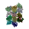
|
|---|---|
| 1 |
|
-Components
| #1: Protein | Mass: 47071.582 Da / Num. of mol.: 8 Source method: isolated from a genetically manipulated source Source: (gene. exp.) Prosthecobacter dejongeii (bacteria) / Gene: btubA / Production host: #2: Protein | Mass: 46465.508 Da / Num. of mol.: 8 Source method: isolated from a genetically manipulated source Source: (gene. exp.) Prosthecobacter dejongeii (bacteria) / Gene: btubB / Production host: #3: Protein | Mass: 27135.703 Da / Num. of mol.: 8 Source method: isolated from a genetically manipulated source Source: (gene. exp.) Prosthecobacter vanneervenii (bacteria)Gene: bklc / Production host: #4: Chemical | ChemComp-GDP /   Type: RNA linking / Mass: 443.201 Da / Num. of mol.: 16 / Source method: obtained synthetically / Formula: C10H15N5O11P2 / Comment: GDP, energy-carrying molecule*YM Type: RNA linking / Mass: 443.201 Da / Num. of mol.: 16 / Source method: obtained synthetically / Formula: C10H15N5O11P2 / Comment: GDP, energy-carrying molecule*YM |
|---|
-Experimental details
-Experiment
| Experiment | Method: ELECTRON MICROSCOPY |
|---|---|
| EM experiment | Aggregation state: HELICAL ARRAY / 3D reconstruction method: helical reconstruction |
- Sample preparation
| Component |
| ||||||||||||||||||||||||
|---|---|---|---|---|---|---|---|---|---|---|---|---|---|---|---|---|---|---|---|---|---|---|---|---|---|
| Molecular weight | Experimental value: NO | ||||||||||||||||||||||||
| Source (natural) |
| ||||||||||||||||||||||||
| Source (recombinant) |
| ||||||||||||||||||||||||
| Buffer solution | pH: 7.7 | ||||||||||||||||||||||||
| Specimen | Embedding applied: NO / Shadowing applied: NO / Staining applied: NO / Vitrification applied: YES | ||||||||||||||||||||||||
| Vitrification | Instrument: FEI VITROBOT MARK IV / Cryogen name: ETHANE |
- Electron microscopy imaging
| Experimental equipment |  Model: Tecnai Polara / Image courtesy: FEI Company |
|---|---|
| Microscopy | Model: FEI POLARA 300 |
| Electron gun | Electron source:  FIELD EMISSION GUN / Accelerating voltage: 300 kV / Illumination mode: FLOOD BEAM FIELD EMISSION GUN / Accelerating voltage: 300 kV / Illumination mode: FLOOD BEAM |
| Electron lens | Mode: BRIGHT FIELD / Alignment procedure: COMA FREE |
| Image recording | Electron dose: 40 e/Å2 / Detector mode: INTEGRATING / Film or detector model: FEI FALCON II (4k x 4k) / Num. of real images: 6105 |
- Processing
| CTF correction | Type: PHASE FLIPPING AND AMPLITUDE CORRECTION |
|---|---|
| Helical symmerty | Angular rotation/subunit: -5.54 ° / Axial rise/subunit: 79.31 Å / Axial symmetry: C1 |
| 3D reconstruction | Resolution: 3.6 Å / Resolution method: FSC 0.143 CUT-OFF / Num. of particles: 257661 / Symmetry type: HELICAL |
| Atomic model building | Protocol: OTHER / Space: RECIPROCAL / Target criteria: R-factor |
